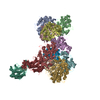
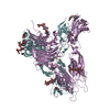
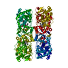
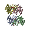
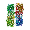
- PDB-5yh2: The structure of DrFam20C1 and hFam20A complex -
+
Open data
ID or keywords:
Loading...
-
Basic information
Entry
Database: PDB / ID: 5yh2
Title
The structure of DrFam20C1 and hFam20A complex
Components
Family with sequence similarity 20, member Ca
Pseudokinase FAM20A
Keywords
TRANSFERASE / complex / kinase
Function / homology
Function and homology information
tooth eruption / biomineral tissue development / enamel mineralization / calcium ion homeostasis / protein serine/threonine kinase activator activity / response to bacterium / Post-translational protein phosphorylation / Regulation of Insulin-like Growth Factor (IGF) transport and uptake by Insulin-like Growth Factor Binding Proteins (IGFBPs) / positive regulation of protein phosphorylation / endoplasmic reticulum ...tooth eruption / biomineral tissue development / enamel mineralization / calcium ion homeostasis / protein serine/threonine kinase activator activity / response to bacterium / Post-translational protein phosphorylation / Regulation of Insulin-like Growth Factor (IGF) transport and uptake by Insulin-like Growth Factor Binding Proteins (IGFBPs) / positive regulation of protein phosphorylation / endoplasmic reticulum / Golgi apparatus / extracellular space / extracellular exosome / ATP binding / metal ion binding / membrane / cytoplasm Similarity search - Function
#262 - Oct 2021 Fifty Years of Open Access to PDB Structures similarity (1)
#29 - May 2002 Penicillin-binding Proteins similarity (1)
#95 - Nov 2007 Multidrug Resistance Transporters similarity (1)
-
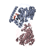
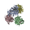
Assembly
Deposited unit
A: Pseudokinase FAM20A C: Family with sequence similarity 20, member Ca B: Pseudokinase FAM20A D: Family with sequence similarity 20, member Ca hetero molecules
In the structure databanks used in Yorodumi, some data are registered as the other names, "COVID-19 virus" and "2019-nCoV". Here are the details of the virus and the list of structure data.
Jan 31, 2019. EMDB accession codes are about to change! (news from PDBe EMDB page)
EMDB accession codes are about to change! (news from PDBe EMDB page)
The allocation of 4 digits for EMDB accession codes will soon come to an end. Whilst these codes will remain in use, new EMDB accession codes will include an additional digit and will expand incrementally as the available range of codes is exhausted. The current 4-digit format prefixed with “EMD-” (i.e. EMD-XXXX) will advance to a 5-digit format (i.e. EMD-XXXXX), and so on. It is currently estimated that the 4-digit codes will be depleted around Spring 2019, at which point the 5-digit format will come into force.
The EM Navigator/Yorodumi systems omit the EMD- prefix.
Related info.:Q: What is EMD? / ID/Accession-code notation in Yorodumi/EM Navigator
Yorodumi is a browser for structure data from EMDB, PDB, SASBDB, etc.
This page is also the successor to EM Navigator detail page, and also detail information page/front-end page for Omokage search.
The word "yorodu" (or yorozu) is an old Japanese word meaning "ten thousand". "mi" (miru) is to see.
Related info.:EMDB / PDB / SASBDB / Comparison of 3 databanks / Yorodumi Search / Aug 31, 2016. New EM Navigator & Yorodumi / Yorodumi Papers / Jmol/JSmol / Function and homology information / Changes in new EM Navigator and Yorodumi
 Movie
Movie Controller


 Open data
 Basic information
 Components
 Keywords
 Function and homology information
 Homo sapiens (human)
 X-RAY DIFFRACTION /
  Authors
 Citation
 Structure visualization
 Downloads & links
 Other downloads


 PDBj
PDBj


 Assembly
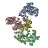
 Trichoplusia ni (cabbage looper) / References: UniProt: Q96MK3
 Mass: 507.181 Da / Num. of mol.: 2
 Sample preparation
 / Beamline: BL18U1 / Wavelength: 0.977 Å
 Processing