 Movie
Movie Controller


+ Open data

- Basic information
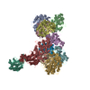
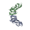
| Entry | Database: PDB / ID: 5yh3 | ||||||
|---|---|---|---|---|---|---|---|
| Title | The structure of hFam20C and hFam20A complex | ||||||
 Components Components |
| ||||||
 Keywords Keywords | TRANSFERASE / complex / kinase | ||||||
| Function / homology |  Function and homology information Function and homology informationosteoclast maturation / regulation of phosphorus metabolic process / tooth eruption / regulation of fibroblast growth factor receptor signaling pathway / biomineral tissue development / dentinogenesis / odontoblast differentiation / enamel mineralization / positive regulation of osteoblast differentiation / positive regulation of bone mineralization ...osteoclast maturation / regulation of phosphorus metabolic process / tooth eruption / regulation of fibroblast growth factor receptor signaling pathway / biomineral tissue development / dentinogenesis / odontoblast differentiation / enamel mineralization / positive regulation of osteoblast differentiation / positive regulation of bone mineralization / calcium ion homeostasis / post-translational protein modification / protein serine/threonine kinase activator activity / response to bacterium / Post-translational protein phosphorylation / positive regulation of protein phosphorylation / Regulation of Insulin-like Growth Factor (IGF) transport and uptake by Insulin-like Growth Factor Binding Proteins (IGFBPs) / manganese ion binding / protease binding / protein phosphorylation / protein kinase activity / non-specific serine/threonine protein kinase / endoplasmic reticulum lumen / Golgi membrane / protein serine kinase activity / protein serine/threonine kinase activity / calcium ion binding / Golgi apparatus / endoplasmic reticulum / : / extracellular exosome / nucleoplasm / ATP binding / cytoplasm Similarity search - Function | ||||||
| Biological species |  Homo sapiens (human) Homo sapiens (human) | ||||||
| Method |  X-RAY DIFFRACTION / SYNCHROTRON / MOLECULAR REPLACEMENT / Resolution: 3.3 Å X-RAY DIFFRACTION / SYNCHROTRON / MOLECULAR REPLACEMENT / Resolution: 3.3 Å | ||||||
 Authors Authors | Zhu, Q. / Xiao, J. | ||||||
 Citation Citation | Journal: Nat Commun / Year: 2018 Title: Structure and evolution of the Fam20 kinases Authors: Zhang, H. / Zhu, Q. / Cui, J. / Wang, Y. / Chen, M.J. / Guo, X. / Tagliabracci, V.S. / Dixon, J.E. / Xiao, J. | ||||||
| History |
|
- Structure visualization
| Structure viewer | Molecule: MolmilJmol/JSmol |
|---|
- Downloads & links
-Download
| PDBx/mmCIF format | 5yh3.cif.gz | 340.2 KB | Display | PDBx/mmCIF format |
|---|---|---|---|---|
| PDB format | pdb5yh3.ent.gz | 267.5 KB | Display | PDB format |
| PDBx/mmJSON format | 5yh3.json.gz | Tree view | PDBx/mmJSON format | |
| Others |  Other downloads Other downloads |
-Validation report
| Arichive directory | https://data.pdbj.org/pub/pdb/validation_reports/yh/5yh3ftp://data.pdbj.org/pub/pdb/validation_reports/yh/5yh3 | HTTPS FTP |
|---|
-Related structure data


-Links
 PDBj
PDBj
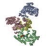
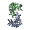
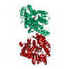
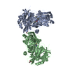
- Assembly
| Deposited unit | 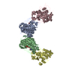
| ||||||||
|---|---|---|---|---|---|---|---|---|---|
| 1 | 
| ||||||||
| 2 | 
| ||||||||
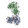
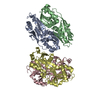
| Unit cell |
|
-Components
| #1: Protein | Mass: 53464.199 Da / Num. of mol.: 2 / Fragment: UNP residues 63-529 Source method: isolated from a genetically manipulated source Source: (gene. exp.) Homo sapiens (human) / Gene: FAM20A, UNQ9388/PRO34279 / Production host:  Trichoplusia ni (cabbage looper) / References: UniProt: Q96MK3 Trichoplusia ni (cabbage looper) / References: UniProt: Q96MK3#2: Protein | Mass: 50683.367 Da / Num. of mol.: 2 / Fragment: UNP residues 141-578 Source method: isolated from a genetically manipulated source Source: (gene. exp.) Homo sapiens (human) / Gene: FAM20C, DMP4 / Production host: Trichoplusia ni (cabbage looper)References: UniProt: Q8IXL6, non-specific serine/threonine protein kinase #3: Chemical | ChemComp-ATP /   Mass: 507.181 Da / Num. of mol.: 4 Mass: 507.181 Da / Num. of mol.: 4Source method: isolated from a genetically manipulated source Formula: C10H16N5O13P3 / Comment: ATP, energy-carrying molecule*YM Has protein modification | Y | |
|---|
-Experimental details
-Experiment
| Experiment | Method: X-RAY DIFFRACTION / Number of used crystals: 1 |
|---|
- Sample preparation
| Crystal | Density Matthews: 4.11 Å3/Da / Density % sol: 70.11 % Description: THE ENTRY CONTAINS FRIEDEL PAIRS IN F_PLUS/MINUS COLUMNS. |
|---|---|
| Crystal grow | Temperature: 290 K / Method: vapor diffusion, sitting drop Details: 0.1M BICINE pH 9.0, 2%(v/v) 1,4-Dioxane, 7%(w/v) Polyethylene glycol 20,000 |
-Data collection
| Diffraction | Mean temperature: 100 K |
|---|---|
| Diffraction source | Source: SYNCHROTRON / Site: SSRF  / Beamline: BL18U1 / Wavelength: 0.977 Å / Beamline: BL18U1 / Wavelength: 0.977 Å |
| Detector | Type: DECTRIS PILATUS3 6M / Detector: PIXEL / Date: Oct 2, 2016 |
| Radiation | Protocol: SINGLE WAVELENGTH / Monochromatic (M) / Laue (L): M / Scattering type: x-ray |
| Radiation wavelength | Wavelength: 0.977 Å / Relative weight: 1 |
| Reflection | Resolution: 3.3→50 Å / Num. obs: 75772 / % possible obs: 99.7 % / Redundancy: 5 % / Net I/σ(I): 10 |
- Processing
| Software |
| ||||||||||||||||||||||||||||||||||||||||||||||||||||||||||||||||||||||||||||||||||||||||||||||||||||||||||||||||||||||||||||||||||||||||||||||||||||||||||||||||||||||||||||||||||||||
|---|---|---|---|---|---|---|---|---|---|---|---|---|---|---|---|---|---|---|---|---|---|---|---|---|---|---|---|---|---|---|---|---|---|---|---|---|---|---|---|---|---|---|---|---|---|---|---|---|---|---|---|---|---|---|---|---|---|---|---|---|---|---|---|---|---|---|---|---|---|---|---|---|---|---|---|---|---|---|---|---|---|---|---|---|---|---|---|---|---|---|---|---|---|---|---|---|---|---|---|---|---|---|---|---|---|---|---|---|---|---|---|---|---|---|---|---|---|---|---|---|---|---|---|---|---|---|---|---|---|---|---|---|---|---|---|---|---|---|---|---|---|---|---|---|---|---|---|---|---|---|---|---|---|---|---|---|---|---|---|---|---|---|---|---|---|---|---|---|---|---|---|---|---|---|---|---|---|---|---|---|---|---|---|
| Refinement | Method to determine structure: MOLECULAR REPLACEMENT / Resolution: 3.3→48.557 Å / SU ML: 0.4 / Cross valid method: FREE R-VALUE / σ(F): 1.34 / Phase error: 26.74 Details: SF FILE CONTAINS FRIEDEL PAIRS UNDER I/F_MINUS AND I/F_PLUS COLUMNS.
| ||||||||||||||||||||||||||||||||||||||||||||||||||||||||||||||||||||||||||||||||||||||||||||||||||||||||||||||||||||||||||||||||||||||||||||||||||||||||||||||||||||||||||||||||||||||
| Solvent computation | Shrinkage radii: 0.9 Å / VDW probe radii: 1.11 Å | ||||||||||||||||||||||||||||||||||||||||||||||||||||||||||||||||||||||||||||||||||||||||||||||||||||||||||||||||||||||||||||||||||||||||||||||||||||||||||||||||||||||||||||||||||||||
| Refinement step | Cycle: LAST / Resolution: 3.3→48.557 Å
| ||||||||||||||||||||||||||||||||||||||||||||||||||||||||||||||||||||||||||||||||||||||||||||||||||||||||||||||||||||||||||||||||||||||||||||||||||||||||||||||||||||||||||||||||||||||
| Refine LS restraints |
| ||||||||||||||||||||||||||||||||||||||||||||||||||||||||||||||||||||||||||||||||||||||||||||||||||||||||||||||||||||||||||||||||||||||||||||||||||||||||||||||||||||||||||||||||||||||
| LS refinement shell |
|
