 Movie
Movie Controller


+ Open data

- Basic information
| Entry | Database: PDB / ID: 5yct | ||||||
|---|---|---|---|---|---|---|---|
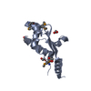
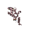
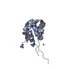
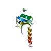
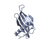
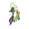
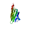
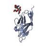
| Title | Engineered hairpin loop3 mutant monomer in Single-chain Monellin | ||||||
 Components Components | Single chain Monellin | ||||||
 Keywords Keywords | PLANT PROTEIN / domain swapped dimer / Single-chain Monellin / loop mutation / QVVAG motif | ||||||
| Function / homology |  Function and homology information Function and homology informationMonellin, B chain / : / Monellin / Monellin / Nuclear Transport Factor 2; Chain: A, - #10 / Cystatin superfamily / Nuclear Transport Factor 2; Chain: A, / Roll / Alpha Beta Similarity search - Domain/homology | ||||||
| Biological species |  Dioscoreophyllum cumminsii (serendipity berry) Dioscoreophyllum cumminsii (serendipity berry) | ||||||
| Method |  X-RAY DIFFRACTION / MOLECULAR REPLACEMENT / Resolution: 1.851 Å X-RAY DIFFRACTION / MOLECULAR REPLACEMENT / Resolution: 1.851 Å | ||||||
 Authors Authors | Surana, P. / Nandwani, N. / Udgaonkar, J.B. / Gosavi, S. / Das, R. | ||||||
 Citation Citation | Journal: Nat Commun / Year: 2019 Title: A five-residue motif for the design of domain swapping in proteins. Authors: Nandwani, N. / Surana, P. / Negi, H. / Mascarenhas, N.M. / Udgaonkar, J.B. / Das, R. / Gosavi, S. | ||||||
| History |
|
- Structure visualization
| Structure viewer | Molecule: MolmilJmol/JSmol |
|---|
- Downloads & links
-Download
| PDBx/mmCIF format | 5yct.cif.gz | 92.3 KB | Display | PDBx/mmCIF format |
|---|---|---|---|---|
| PDB format | pdb5yct.ent.gz | 69.6 KB | Display | PDB format |
| PDBx/mmJSON format | 5yct.json.gz | Tree view | PDBx/mmJSON format | |
| Others |  Other downloads Other downloads |
-Validation report
| Arichive directory | https://data.pdbj.org/pub/pdb/validation_reports/yc/5yctftp://data.pdbj.org/pub/pdb/validation_reports/yc/5yct | HTTPS FTP |
|---|
-Related structure data
| Related structure data |  5ycuC  5ycwC 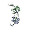 6iwjC  1iv7S S: Starting model for refinement C: citing same article ( |
|---|---|
| Similar structure data |


-Links
 PDBj
PDBj
- Assembly
| Deposited unit | 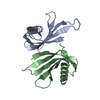
| ||||||||
|---|---|---|---|---|---|---|---|---|---|
| 1 | 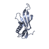
| ||||||||
| 2 | 
| ||||||||
| Unit cell |
|
-Components
| #1: Protein | Mass: 11493.126 Da / Num. of mol.: 2 / Mutation: DYKTR to QVVAG in loop3 Source method: isolated from a genetically manipulated source Source: (gene. exp.) Dioscoreophyllum cumminsii (serendipity berry)Production host:  #2: Water | ChemComp-HOH / |  Mass: 18.015 Da / Num. of mol.: 154 / Source method: isolated from a natural source / Formula: H2O Mass: 18.015 Da / Num. of mol.: 154 / Source method: isolated from a natural source / Formula: H2OSequence details | The complete sequence of single chain Monellin has been deposited to NCBI with accession code ...The complete sequence of single chain Monellin has been deposited to NCBI with accession code AFF58925. Residues 79-83 DYKTR have been replaced with QVVAG in this structure. C-terminal residues STP are from expression tag. | |
|---|
-Experimental details
-Experiment
| Experiment | Method: X-RAY DIFFRACTION / Number of used crystals: 1 |
|---|
- Sample preparation
| Crystal | Density Matthews: 2.27 Å3/Da / Density % sol: 45.81 % |
|---|---|
| Crystal grow | Temperature: 277 K / Method: vapor diffusion, hanging drop Details: 8-12% (wt/vol) PEG 8000, 50mM sodium phosphate, pH 6.4-7.2, Crystals grew to maximum size in 4-5 days PH range: 6.4-7.2 |
-Data collection
| Diffraction | Mean temperature: 100 K |
|---|---|
| Diffraction source | Source: ROTATING ANODE / Type: RIGAKU FR-X / Wavelength: 1.5418 Å |
| Detector | Type: RIGAKU RAXIS IV++ / Detector: IMAGE PLATE / Date: Mar 16, 2017 |
| Radiation | Protocol: SINGLE WAVELENGTH / Monochromatic (M) / Laue (L): M / Scattering type: x-ray |
| Radiation wavelength | Wavelength: 1.5418 Å / Relative weight: 1 |
| Reflection | Resolution: 1.851→24.052 Å / Num. obs: 17514 / % possible obs: 98.8 % / Redundancy: 4.8 % / Biso Wilson estimate: 32.3 Å2 / CC1/2: 0.999 / Rmerge(I) obs: 0.017 / Net I/σ(I): 24 |
| Reflection shell | Highest resolution: 1.851 Å / Rmerge(I) obs: 0.195 / Mean I/σ(I) obs: 3.1 / CC1/2: 0.924 / % possible all: 89.04 |
- Processing
| Software |
| |||||||||||||||||||||||||||||||||||||||||||||||||||||||||||||||||||||||||||
|---|---|---|---|---|---|---|---|---|---|---|---|---|---|---|---|---|---|---|---|---|---|---|---|---|---|---|---|---|---|---|---|---|---|---|---|---|---|---|---|---|---|---|---|---|---|---|---|---|---|---|---|---|---|---|---|---|---|---|---|---|---|---|---|---|---|---|---|---|---|---|---|---|---|---|---|---|
| Refinement | Method to determine structure: MOLECULAR REPLACEMENT Starting model: 1IV7 Resolution: 1.851→24.052 Å / SU ML: 0.21 / Cross valid method: FREE R-VALUE / σ(F): 1.36 / Phase error: 25.93
| |||||||||||||||||||||||||||||||||||||||||||||||||||||||||||||||||||||||||||
| Solvent computation | Shrinkage radii: 0.9 Å / VDW probe radii: 1.11 Å | |||||||||||||||||||||||||||||||||||||||||||||||||||||||||||||||||||||||||||
| Refinement step | Cycle: LAST / Resolution: 1.851→24.052 Å
| |||||||||||||||||||||||||||||||||||||||||||||||||||||||||||||||||||||||||||
| Refine LS restraints |
| |||||||||||||||||||||||||||||||||||||||||||||||||||||||||||||||||||||||||||
| LS refinement shell |
| |||||||||||||||||||||||||||||||||||||||||||||||||||||||||||||||||||||||||||
| Refinement TLS params. | Method: refined / Refine-ID: X-RAY DIFFRACTION
| |||||||||||||||||||||||||||||||||||||||||||||||||||||||||||||||||||||||||||
| Refinement TLS group |
|
