 Movie
Movie Controller


+ Open data

- Basic information
| Entry | Database: PDB / ID: 5y31 | ||||||
|---|---|---|---|---|---|---|---|
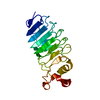
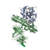
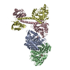
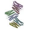
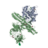
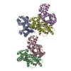
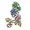
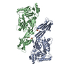
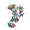
| Title | Crystal structure of human LGI1-ADAM22 complex | ||||||
 Components Components |
| ||||||
 Keywords Keywords | CELL ADHESION / epilepsy / synapse / ADAM / EPTP / WD40 | ||||||
| Function / homology |  Function and homology information Function and homology informationLGI-ADAM interactions / negative regulation of cell adhesion / axon initial segment / neurotransmitter receptor localization to postsynaptic specialization membrane / positive regulation of synaptic transmission / synaptic cleft / axon guidance / central nervous system development / metalloendopeptidase activity / postsynaptic density membrane ...LGI-ADAM interactions / negative regulation of cell adhesion / axon initial segment / neurotransmitter receptor localization to postsynaptic specialization membrane / positive regulation of synaptic transmission / synaptic cleft / axon guidance / central nervous system development / metalloendopeptidase activity / postsynaptic density membrane / integrin binding / neuron projection development / nervous system development / signaling receptor activity / positive regulation of cell growth / cell adhesion / receptor ligand activity / signaling receptor binding / axon / dendrite / glutamatergic synapse / Golgi apparatus / endoplasmic reticulum / proteolysis / : / extracellular region / membrane / plasma membrane / cytoplasm Similarity search - Function | ||||||
| Biological species |  Homo sapiens (human) Homo sapiens (human) | ||||||
| Method |  X-RAY DIFFRACTION / SYNCHROTRON / MOLECULAR REPLACEMENT / Resolution: 7.125 Å X-RAY DIFFRACTION / SYNCHROTRON / MOLECULAR REPLACEMENT / Resolution: 7.125 Å | ||||||
 Authors Authors | Yamagata, A. / Fukai, S. | ||||||
 Citation Citation | Journal: Nat Commun / Year: 2018 Title: Structural basis of epilepsy-related ligand-receptor complex LGI1-ADAM22. Authors: Yamagata, A. / Miyazaki, Y. / Yokoi, N. / Shigematsu, H. / Sato, Y. / Goto-Ito, S. / Maeda, A. / Goto, T. / Sanbo, M. / Hirabayashi, M. / Shirouzu, M. / Fukata, Y. / Fukata, M. / Fukai, S. | ||||||
| History |
|
- Structure visualization
| Structure viewer | Molecule: MolmilJmol/JSmol |
|---|
- Downloads & links
-Download
| PDBx/mmCIF format | 5y31.cif.gz | 819.8 KB | Display | PDBx/mmCIF format |
|---|---|---|---|---|
| PDB format | pdb5y31.ent.gz | 685.3 KB | Display | PDB format |
| PDBx/mmJSON format | 5y31.json.gz | Tree view | PDBx/mmJSON format | |
| Others |  Other downloads Other downloads |
-Validation report
| Arichive directory | https://data.pdbj.org/pub/pdb/validation_reports/y3/5y31ftp://data.pdbj.org/pub/pdb/validation_reports/y3/5y31 | HTTPS FTP |
|---|
-Related structure data

-Links
 PDBj
PDBj


- Assembly
| Deposited unit | 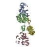
| ||||||||
|---|---|---|---|---|---|---|---|---|---|
| 1 | 
| ||||||||
| 2 | 
| ||||||||
| Unit cell |
|
-Components
| #1: Protein | Mass: 54803.230 Da / Num. of mol.: 2 / Fragment: UNP RESIDUES 233-729 Source method: isolated from a genetically manipulated source Source: (gene. exp.) Homo sapiens (human) / Gene: ADAM22, MDC2 / Production host: Mammalia (mammals) / References: UniProt: Q9P0K1#2: Protein | Mass: 62570.785 Da / Num. of mol.: 2 / Fragment: UNP RESIDUES 37-557 / Mutation: R470A Source method: isolated from a genetically manipulated source Source: (gene. exp.) Homo sapiens (human) / Gene: LGI1, EPT, UNQ775/PRO1569 / Production host: Mammalia (mammals) / References: UniProt: O95970#3: Chemical | ChemComp-CA /   Mass: 40.078 Da / Num. of mol.: 8 Mass: 40.078 Da / Num. of mol.: 8Source method: isolated from a genetically manipulated source Formula: Ca #4: Sugar | ChemComp-NAG /   Type: D-saccharide, beta linking / Mass: 221.208 Da / Num. of mol.: 10 Type: D-saccharide, beta linking / Mass: 221.208 Da / Num. of mol.: 10Source method: isolated from a genetically manipulated source Formula: C8H15NO6 Has protein modification | Y | |
|---|
-Experimental details
-Experiment
| Experiment | Method: X-RAY DIFFRACTION / Number of used crystals: 1 |
|---|
- Sample preparation
| Crystal | Density Matthews: 4.39 Å3/Da / Density % sol: 71.99 % |
|---|---|
| Crystal grow | Temperature: 293 K / Method: vapor diffusion, sitting drop / pH: 6 Details: 10 % PEG 8000, 0.1 M zinc acetate, 0.1 M MES-Na (pH 6.0) |
-Data collection
| Diffraction | Mean temperature: 100 K |
|---|---|
| Diffraction source | Source: SYNCHROTRON / Site: SPring-8  / Beamline: BL41XU / Wavelength: 1 Å / Beamline: BL41XU / Wavelength: 1 Å |
| Detector | Type: DECTRIS PILATUS3 6M / Detector: PIXEL / Date: Apr 15, 2016 |
| Radiation | Protocol: SINGLE WAVELENGTH / Monochromatic (M) / Laue (L): M / Scattering type: x-ray |
| Radiation wavelength | Wavelength: 1 Å / Relative weight: 1 |
| Reflection | Resolution: 7.12→50 Å / Num. obs: 6092 / % possible obs: 97.9 % / Redundancy: 7.1 % / Net I/σ(I): 13.3 |
| Reflection shell | Resolution: 7.12→7.24 Å / Redundancy: 4.6 % / Mean I/σ(I) obs: 4.6 / CC1/2: 0.578 / Rsym value: 0.461 / % possible all: 96.3 |
- Processing
| Software |
| ||||||||||||||||||||||||||||||||||||||||
|---|---|---|---|---|---|---|---|---|---|---|---|---|---|---|---|---|---|---|---|---|---|---|---|---|---|---|---|---|---|---|---|---|---|---|---|---|---|---|---|---|---|
| Refinement | Method to determine structure: MOLECULAR REPLACEMENT / Resolution: 7.125→48.993 Å / SU ML: 1.37 / Cross valid method: FREE R-VALUE / σ(F): 1.45 / Phase error: 37.83 / Stereochemistry target values: ML
| ||||||||||||||||||||||||||||||||||||||||
| Solvent computation | Shrinkage radii: 0.9 Å / VDW probe radii: 1.11 Å / Solvent model: FLAT BULK SOLVENT MODEL | ||||||||||||||||||||||||||||||||||||||||
| Refinement step | Cycle: LAST / Resolution: 7.125→48.993 Å
| ||||||||||||||||||||||||||||||||||||||||
| Refine LS restraints |
| ||||||||||||||||||||||||||||||||||||||||
| LS refinement shell |
| ||||||||||||||||||||||||||||||||||||||||
| Refinement TLS params. | Method: refined / Origin x: 2.1416 Å / Origin y: -1.187 Å / Origin z: 51.1431 Å
| ||||||||||||||||||||||||||||||||||||||||
| Refinement TLS group | Selection details: all |
