 Movie
Movie Controller

[English] 日本語
 Yorodumi
- PDB-5xrn: Galectin-10/Charcot-Leyden crystal protein variant K73A crystal s... -
+ Open data

- Basic information
| Entry | Database: PDB / ID: 5xrn | ||||||
|---|---|---|---|---|---|---|---|
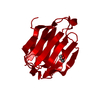
| Title | Galectin-10/Charcot-Leyden crystal protein variant K73A crystal structure | ||||||
 Components Components | Galectin-10 | ||||||
 Keywords Keywords | PROTEIN BINDING / Galectin-10/Charcot-Leyden crystal protein | ||||||
| Function / homology |  Function and homology information Function and homology informationregulation of activated T cell proliferation / regulation of T cell cytokine production / T cell apoptotic process / regulation of T cell anergy / carbohydrate binding / extracellular matrix / identical protein binding / cytosol Similarity search - Function | ||||||
| Biological species |  Homo sapiens (human) Homo sapiens (human) | ||||||
| Method |  X-RAY DIFFRACTION / SYNCHROTRON / Resolution: 1.6 Å X-RAY DIFFRACTION / SYNCHROTRON / Resolution: 1.6 Å | ||||||
 Authors Authors | Su, J. | ||||||
 Citation Citation | Journal: Glycobiology / Year: 2018 Title: Galectin-10: a new structural type of prototype galectin dimer and effects on saccharide ligand binding. Authors: Su, J. / Gao, J. / Si, Y. / Cui, L. / Song, C. / Wang, Y. / Wu, R. / Tai, G. / Zhou, Y. | ||||||
| History |
|
- Structure visualization
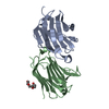
| Structure viewer | Molecule: MolmilJmol/JSmol |
|---|
- Downloads & links
-Download
| PDBx/mmCIF format | 5xrn.cif.gz | 46 KB | Display | PDBx/mmCIF format |
|---|---|---|---|---|
| PDB format | pdb5xrn.ent.gz | 30.8 KB | Display | PDB format |
| PDBx/mmJSON format | 5xrn.json.gz | Tree view | PDBx/mmJSON format | |
| Others |  Other downloads Other downloads |
-Validation report
| Arichive directory | https://data.pdbj.org/pub/pdb/validation_reports/xr/5xrnftp://data.pdbj.org/pub/pdb/validation_reports/xr/5xrn | HTTPS FTP |
|---|
-Related structure data
| Related structure data |  5xrgC  5xrhC  5xriC  5xrjC  5xrkC  5xrlC  5xrmC  5xroC  5xrpC  5yt4C C: citing same article ( |
|---|---|
| Similar structure data |


-Links
 PDBj
PDBj- Assembly
| Deposited unit | 
| |||||||||
|---|---|---|---|---|---|---|---|---|---|---|
| 1 | 
| |||||||||
| Unit cell |
| |||||||||
| Components on special symmetry positions |
|
-Components
| #1: Protein | Mass: 16696.006 Da / Num. of mol.: 1 / Mutation: K73A Source method: isolated from a genetically manipulated source Source: (gene. exp.) Homo sapiens (human) / Gene: CLC, LGALS10, LGALS10A / Production host:  |
|---|---|
| #2: Chemical | ChemComp-GOL /   Mass: 92.094 Da / Num. of mol.: 1 / Source method: obtained synthetically / Formula: C3H8O3 Mass: 92.094 Da / Num. of mol.: 1 / Source method: obtained synthetically / Formula: C3H8O3 |
| #3: Water | ChemComp-HOH /  Mass: 18.015 Da / Num. of mol.: 171 / Source method: isolated from a natural source / Formula: H2O Mass: 18.015 Da / Num. of mol.: 171 / Source method: isolated from a natural source / Formula: H2O |
-Experimental details
-Experiment
| Experiment | Method: X-RAY DIFFRACTION / Number of used crystals: 1 |
|---|
- Sample preparation
| Crystal | Density Matthews: 2.75 Å3/Da / Density % sol: 55.34 % |
|---|---|
| Crystal grow | Temperature: 298 K / Method: vapor diffusion, hanging drop / Details: Bis-Tris |
-Data collection
| Diffraction | Mean temperature: 100 K |
|---|---|
| Diffraction source | Source: SYNCHROTRON / Site: SSRF  / Beamline: BL18U1 / Wavelength: 0.9 Å / Beamline: BL18U1 / Wavelength: 0.9 Å |
| Detector | Type: DECTRIS PILATUS3 6M / Detector: PIXEL / Date: Mar 19, 2017 |
| Radiation | Protocol: SINGLE WAVELENGTH / Monochromatic (M) / Laue (L): M / Scattering type: x-ray |
| Radiation wavelength | Wavelength: 0.9 Å / Relative weight: 1 |
| Reflection | Resolution: 1.6→19.624 Å / Num. obs: 25733 / % possible obs: 99.9 % / Redundancy: 18.4 % / Net I/σ(I): 14.5 |
- Processing
| Software |
| ||||||||||||||||||||||||
|---|---|---|---|---|---|---|---|---|---|---|---|---|---|---|---|---|---|---|---|---|---|---|---|---|---|
| Refinement | Resolution: 1.6→19.624 Å / SU ML: 0.14 / Cross valid method: NONE / σ(F): 1.37 / Phase error: 18.17
| ||||||||||||||||||||||||
| Solvent computation | Shrinkage radii: 0.9 Å / VDW probe radii: 1.11 Å | ||||||||||||||||||||||||
| Displacement parameters | Biso max: 50.49 Å2 / Biso mean: 19.3365 Å2 / Biso min: 8.12 Å2 | ||||||||||||||||||||||||
| Refinement step | Cycle: final / Resolution: 1.6→19.624 Å
|
