 Movie
Movie Controller


+ Open data

- Basic information
| Entry | Database: PDB / ID: 5xm6 | ||||||
|---|---|---|---|---|---|---|---|
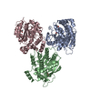
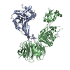
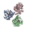
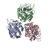
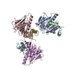
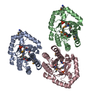
| Title | the overall structure of VrEH2 | ||||||
 Components Components | Epoxide hydrolase | ||||||
 Keywords Keywords | HYDROLASE / epoxide hydrolase | ||||||
| Function / homology |  Function and homology information Function and homology informationHydrolases; Acting on ether bonds; Ether hydrolases / soluble epoxide hydrolase / epoxide hydrolase activity Similarity search - Function | ||||||
| Biological species |  Vigna radiata (mung bean) Vigna radiata (mung bean) | ||||||
| Method |  X-RAY DIFFRACTION / SYNCHROTRON / MOLECULAR REPLACEMENT / Resolution: 2.501 Å X-RAY DIFFRACTION / SYNCHROTRON / MOLECULAR REPLACEMENT / Resolution: 2.501 Å | ||||||
 Authors Authors | Li, F.L. / Kong, X.D. / Yu, H.L. / Shang, Y.P. / Zhou, J.H. / Xu, J.H. | ||||||
 Citation Citation | Journal: Acs Catalysis / Year: 2018 Title: Regioselectivity Engineering of Epoxide Hydrolase: Near-Perfect Enantioconvergence through a Single Site Mutation Authors: Li, F.L. / Kong, X.D. / Chen, Q. / Zheng, Y.C. / Xu, Q. / Chen, F.F. / Fan, L.Q. / Lin, G.Q. / Zhou, J.H. / Yu, H.L. / Xu, J.H. | ||||||
| History |
|
- Structure visualization
| Structure viewer | Molecule: MolmilJmol/JSmol |
|---|
- Downloads & links
-Download
| PDBx/mmCIF format | 5xm6.cif.gz | 212.2 KB | Display | PDBx/mmCIF format |
|---|---|---|---|---|
| PDB format | pdb5xm6.ent.gz | 168.2 KB | Display | PDB format |
| PDBx/mmJSON format | 5xm6.json.gz | Tree view | PDBx/mmJSON format | |
| Others |  Other downloads Other downloads |
-Validation report
| Arichive directory | https://data.pdbj.org/pub/pdb/validation_reports/xm/5xm6ftp://data.pdbj.org/pub/pdb/validation_reports/xm/5xm6 | HTTPS FTP |
|---|
-Related structure data
| Related structure data | 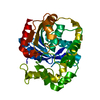 5y6yC 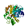 5yb5C 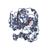 2cjpS S: Starting model for refinement C: citing same article ( |
|---|---|
| Similar structure data |

-Links
 PDBj
PDBj


- Assembly
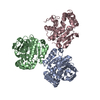
| Deposited unit | 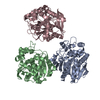
| ||||||||
|---|---|---|---|---|---|---|---|---|---|
| 1 |
| ||||||||
| Unit cell |
|
-Components
| #1: Protein | Mass: 39815.141 Da / Num. of mol.: 3 / Mutation: G3E, V4I, R114H Source method: isolated from a genetically manipulated source Source: (gene. exp.) Vigna radiata (mung bean) / Gene: Vreh3 / Production host:  References: UniProt: A0A0G3F3K2, UniProt: A0A0R5NGA4*PLUS, Hydrolases; Acting on ether bonds; Ether hydrolases #2: Water | ChemComp-HOH / |  Mass: 18.015 Da / Num. of mol.: 572 / Source method: isolated from a natural source / Formula: H2O Mass: 18.015 Da / Num. of mol.: 572 / Source method: isolated from a natural source / Formula: H2O |
|---|
-Experimental details
-Experiment
| Experiment | Method: X-RAY DIFFRACTION / Number of used crystals: 1 |
|---|
- Sample preparation
| Crystal | Density Matthews: 3.25 Å3/Da / Density % sol: 62.1 % / Description: rhombus |
|---|---|
| Crystal grow | Temperature: 285 K / Method: vapor diffusion, sitting drop / pH: 7.5 Details: PEG 20000, PEG500MME, HEPES, Mops, sodium nitrate, ammonium sulfate, disodium hydrogen phosphate. |
-Data collection
| Diffraction | Mean temperature: 100 K |
|---|---|
| Diffraction source | Source: SYNCHROTRON / Site: SSRF  / Beamline: BL17U1 / Wavelength: 0.9791 Å / Beamline: BL17U1 / Wavelength: 0.9791 Å |
| Detector | Type: ADSC QUANTUM 315r / Detector: CCD / Date: Apr 27, 2016 |
| Radiation | Protocol: SINGLE WAVELENGTH / Monochromatic (M) / Laue (L): M / Scattering type: x-ray |
| Radiation wavelength | Wavelength: 0.9791 Å / Relative weight: 1 |
| Reflection | Resolution: 2.5→50 Å / Num. obs: 50435 / % possible obs: 100 % / Redundancy: 27.9 % / CC1/2: 0.987 / Rpim(I) all: 0.031 / Χ2: 1.022 / Net I/σ(I): 8.25 |
| Reflection shell | Resolution: 2.5→2.59 Å / Redundancy: 29.5 % / Num. unique obs: 4938 / CC1/2: 0.987 / % possible all: 100 |
- Processing
| Software |
| |||||||||||||||||||||||||||||||||||||||||||||||||||||||||||||||||||||||||||||||||||||||||||||||||||||||||||||||||||||||||||||||||||||
|---|---|---|---|---|---|---|---|---|---|---|---|---|---|---|---|---|---|---|---|---|---|---|---|---|---|---|---|---|---|---|---|---|---|---|---|---|---|---|---|---|---|---|---|---|---|---|---|---|---|---|---|---|---|---|---|---|---|---|---|---|---|---|---|---|---|---|---|---|---|---|---|---|---|---|---|---|---|---|---|---|---|---|---|---|---|---|---|---|---|---|---|---|---|---|---|---|---|---|---|---|---|---|---|---|---|---|---|---|---|---|---|---|---|---|---|---|---|---|---|---|---|---|---|---|---|---|---|---|---|---|---|---|---|---|
| Refinement | Method to determine structure: MOLECULAR REPLACEMENT Starting model: 2CJP Resolution: 2.501→41.532 Å / SU ML: 0.27 / Cross valid method: FREE R-VALUE / σ(F): 1.34 / Phase error: 21.78
| |||||||||||||||||||||||||||||||||||||||||||||||||||||||||||||||||||||||||||||||||||||||||||||||||||||||||||||||||||||||||||||||||||||
| Solvent computation | Shrinkage radii: 0.9 Å / VDW probe radii: 1.11 Å | |||||||||||||||||||||||||||||||||||||||||||||||||||||||||||||||||||||||||||||||||||||||||||||||||||||||||||||||||||||||||||||||||||||
| Refinement step | Cycle: LAST / Resolution: 2.501→41.532 Å
| |||||||||||||||||||||||||||||||||||||||||||||||||||||||||||||||||||||||||||||||||||||||||||||||||||||||||||||||||||||||||||||||||||||
| Refine LS restraints |
| |||||||||||||||||||||||||||||||||||||||||||||||||||||||||||||||||||||||||||||||||||||||||||||||||||||||||||||||||||||||||||||||||||||
| LS refinement shell |
|
