 Movie
Movie Controller

[English] 日本語
 Yorodumi
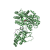
- PDB-5xe3: Endoribonuclease in complex with its cognate antitoxin from Mycob... -
+ Open data

- Basic information
| Entry | Database: PDB / ID: 5xe3 | |||||||||
|---|---|---|---|---|---|---|---|---|---|---|
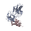
| Title | Endoribonuclease in complex with its cognate antitoxin from Mycobacterial species | |||||||||
 Components Components |
| |||||||||
 Keywords Keywords | HYDROLASE/ANTITOXIN / endonuclease / HYDROLASE-ANTITOXIN complex | |||||||||
| Function / homology |  Function and homology information Function and homology informationrRNA catabolic process / mRNA catabolic process / RNA endonuclease activity / enzyme inhibitor activity / Hydrolases; Acting on ester bonds / hydrolase activity / DNA binding Similarity search - Function | |||||||||
| Biological species |   Mycobacterium tuberculosis (bacteria) Mycobacterium tuberculosis (bacteria) | |||||||||
| Method |  X-RAY DIFFRACTION / SYNCHROTRON / MOLECULAR REPLACEMENT / Resolution: 2.3 Å X-RAY DIFFRACTION / SYNCHROTRON / MOLECULAR REPLACEMENT / Resolution: 2.3 Å | |||||||||
 Authors Authors | Ahn, D.-H. / Lee, K.-Y. / Lee, S.J. / Yoon, H.J. / Kim, S.-J. / Lee, B.-J. | |||||||||
| Funding support |  Korea, Republic Of, 2items Korea, Republic Of, 2items
| |||||||||
 Citation Citation | Journal: J. Biol. Chem. / Year: 2017 Title: Structural analyses of the MazEF4 toxin-antitoxin pair in Mycobacterium tuberculosis provide evidence for a unique extracellular death factor. Authors: Ahn, D.H. / Lee, K.Y. / Lee, S.J. / Park, S.J. / Yoon, H.J. / Kim, S.J. / Lee, B.J. | |||||||||
| History |
|
- Structure visualization
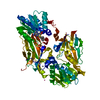
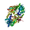
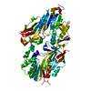
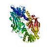
| Structure viewer | Molecule: MolmilJmol/JSmol |
|---|
- Downloads & links
-Download
| PDBx/mmCIF format | 5xe3.cif.gz | 214.6 KB | Display | PDBx/mmCIF format |
|---|---|---|---|---|
| PDB format | pdb5xe3.ent.gz | 174.3 KB | Display | PDB format |
| PDBx/mmJSON format | 5xe3.json.gz | Tree view | PDBx/mmJSON format | |
| Others |  Other downloads Other downloads |
-Validation report
| Arichive directory | https://data.pdbj.org/pub/pdb/validation_reports/xe/5xe3ftp://data.pdbj.org/pub/pdb/validation_reports/xe/5xe3 | HTTPS FTP |
|---|
-Related structure data
| Related structure data |  5xe2C 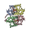 3se5S C: citing same article ( S: Starting model for refinement |
|---|---|
| Similar structure data |


-Links
 PDBj
PDBj- Assembly
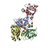
| Deposited unit | 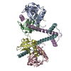
| ||||||||
|---|---|---|---|---|---|---|---|---|---|
| 1 |
| ||||||||
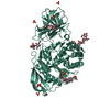
| Unit cell |
|
-Components
| #1: Protein | Mass: 11697.274 Da / Num. of mol.: 4 Source method: isolated from a genetically manipulated source Source: (gene. exp.) Mycobacterium tuberculosis (strain ATCC 25618 / H37Rv) (bacteria)Strain: ATCC 25618 / H37Rv / Gene: mazF4, mazF-mt7, Rv1495, MTCY277.17 / Production host: References: UniProt: P9WII5, Hydrolases; Acting on ester bonds #2: Protein | Mass: 9211.135 Da / Num. of mol.: 2 / Fragment: UNP RESIDUES 19-99 Source method: isolated from a genetically manipulated source Source: (gene. exp.) Mycobacterium tuberculosis (strain ATCC 25618 / H37Rv) (bacteria)Strain: ATCC 25618 / H37Rv / Gene: mazE4, mazE-mt7, Rv1494, MTCY277.16 / Production host: #3: Water | ChemComp-HOH / |  Mass: 18.015 Da / Num. of mol.: 70 / Source method: isolated from a natural source / Formula: H2O Mass: 18.015 Da / Num. of mol.: 70 / Source method: isolated from a natural source / Formula: H2O |
|---|
-Experimental details
-Experiment
| Experiment | Method: X-RAY DIFFRACTION / Number of used crystals: 1 |
|---|
- Sample preparation
| Crystal | Density Matthews: 2.37 Å3/Da / Density % sol: 48.04 % |
|---|---|
| Crystal grow | Temperature: 293 K / Method: evaporation / pH: 7 Details: 0.2 M calcium acetate, 0.1 M Tris-HCl pH 7.0, 20% (w/v) polyethylene glycol 3000 |
-Data collection
| Diffraction | Mean temperature: 100 K |
|---|---|
| Diffraction source | Source: SYNCHROTRON / Site: PAL/PLS / Beamline: 5C (4A) / Wavelength: 0.97945 Å |
| Detector | Type: ADSC QUANTUM 315r / Detector: CCD / Date: Mar 20, 2015 |
| Radiation | Protocol: SINGLE WAVELENGTH / Monochromatic (M) / Laue (L): M / Scattering type: x-ray |
| Radiation wavelength | Wavelength: 0.97945 Å / Relative weight: 1 |
| Reflection | Resolution: 2.3→30 Å / Num. obs: 23785 / % possible obs: 95.21 % / Redundancy: 4 % / Net I/σ(I): 30.21 |
- Processing
| Software |
| ||||||||||||||||||||||||||||||||||||||||||||||||||||||||||||
|---|---|---|---|---|---|---|---|---|---|---|---|---|---|---|---|---|---|---|---|---|---|---|---|---|---|---|---|---|---|---|---|---|---|---|---|---|---|---|---|---|---|---|---|---|---|---|---|---|---|---|---|---|---|---|---|---|---|---|---|---|---|
| Refinement | Method to determine structure: MOLECULAR REPLACEMENT Starting model: 3SE5 Resolution: 2.3→30 Å / Cor.coef. Fo:Fc: 0.943 / Cor.coef. Fo:Fc free: 0.908 / SU B: 9.27 / SU ML: 0.22 / Cross valid method: THROUGHOUT / σ(F): 0 / ESU R: 0.434 / ESU R Free: 0.274 Details: HYDROGENS HAVE BEEN ADDED IN THE RIDING POSITIONS U VALUES
| ||||||||||||||||||||||||||||||||||||||||||||||||||||||||||||
| Solvent computation | Ion probe radii: 0.8 Å / Shrinkage radii: 0.8 Å / VDW probe radii: 1.2 Å | ||||||||||||||||||||||||||||||||||||||||||||||||||||||||||||
| Displacement parameters | Biso max: 121.57 Å2 / Biso mean: 44.244 Å2 / Biso min: 22.49 Å2
| ||||||||||||||||||||||||||||||||||||||||||||||||||||||||||||
| Refinement step | Cycle: final / Resolution: 2.3→30 Å
| ||||||||||||||||||||||||||||||||||||||||||||||||||||||||||||
| Refine LS restraints |
| ||||||||||||||||||||||||||||||||||||||||||||||||||||||||||||
| LS refinement shell | Resolution: 2.301→2.36 Å / Rfactor Rfree error: 0 / Total num. of bins used: 20
|
