Protocol: SINGLE WAVELENGTH / Monochromatic (M) / Laue (L): M / Scattering type: x-ray
Radiation wavelength
Wavelength: 0.979 Å / Relative weight: 1
Reflection
Resolution: 1.6→30 Å / Num. obs: 107019 / % possible obs: 99.9 % / Observed criterion σ(I): 0.5 / Redundancy: 8.7 % / Biso Wilson estimate: 28.86 Å2 / Rmerge(I) obs: 0.08 / Net I/σ(I): 13.9
Reflection shell
Resolution: 1.6→1.63 Å / Redundancy: 8.4 % / Mean I/σ(I) obs: 0.5 / % possible all: 99.9
-
Processing
Software
Name
Version
Classification
PHENIX
(PHENIX.REFINE)
refinement
XDS
datareduction
Aimless
datascaling
PHASER
phasing
Refinement
Method to determine structure: MOLECULAR REPLACEMENT Starting model: SAME STRUCTURE SOLVED BY SAD USING A CRYSTAL OF THE NATIVE PROTEIN SOAKED WITH THIMEROSAL AND PHASED WITH ANOMALOUS SIGNAL FROM MERCURY Resolution: 1.7→29.353 Å / SU ML: 0.21 / σ(F): 1.36 / Phase error: 19.12 / Stereochemistry target values: ML
Rfactor
Num. reflection
% reflection
Rfree
0.1897
4438
5 %
Rwork
0.1597
-
-
obs
0.1612
89408
99.83 %
Solvent computation
Shrinkage radii: 0.9 Å / VDW probe radii: 1.11 Å / Solvent model: FLAT BULK SOLVENT MODEL
Refinement step
Cycle: LAST / Resolution: 1.7→29.353 Å
Protein
Nucleic acid
Ligand
Solvent
Total
Num. atoms
5093
0
52
692
5837
Refine LS restraints
Refine-ID
Type
Dev ideal
Number
X-RAY DIFFRACTION
f_bond_d
0.007
5334
X-RAY DIFFRACTION
f_angle_d
1.131
7265
X-RAY DIFFRACTION
f_dihedral_angle_d
14.082
1974
X-RAY DIFFRACTION
f_chiral_restr
0.045
795
X-RAY DIFFRACTION
f_plane_restr
0.005
935
LS refinement shell
Resolution (Å)
Rfactor Rfree
Num. reflection Rfree
Rfactor Rwork
Num. reflection Rwork
Refine-ID
% reflection obs (%)
1.7-1.7193
0.3523
136
0.3251
2792
X-RAY DIFFRACTION
100
1.7193-1.7395
0.324
151
0.3159
2813
X-RAY DIFFRACTION
100
1.7395-1.7608
0.333
140
0.2916
2757
X-RAY DIFFRACTION
100
1.7608-1.783
0.304
130
0.2716
2825
X-RAY DIFFRACTION
100
1.783-1.8065
0.2912
141
0.2538
2785
X-RAY DIFFRACTION
100
1.8065-1.8312
0.2645
122
0.2216
2834
X-RAY DIFFRACTION
100
1.8312-1.8574
0.2522
167
0.2174
2779
X-RAY DIFFRACTION
100
1.8574-1.8851
0.2271
160
0.2001
2785
X-RAY DIFFRACTION
100
1.8851-1.9146
0.2295
146
0.1908
2797
X-RAY DIFFRACTION
100
1.9146-1.946
0.2327
144
0.1853
2812
X-RAY DIFFRACTION
100
1.946-1.9795
0.2075
147
0.1829
2803
X-RAY DIFFRACTION
100
1.9795-2.0155
0.2216
156
0.1826
2788
X-RAY DIFFRACTION
100
2.0155-2.0542
0.2007
145
0.1719
2840
X-RAY DIFFRACTION
100
2.0542-2.0962
0.193
146
0.1692
2786
X-RAY DIFFRACTION
100
2.0962-2.1417
0.219
148
0.1592
2821
X-RAY DIFFRACTION
100
2.1417-2.1915
0.217
149
0.1624
2791
X-RAY DIFFRACTION
100
2.1915-2.2463
0.184
147
0.1536
2815
X-RAY DIFFRACTION
100
2.2463-2.307
0.1914
150
0.1528
2842
X-RAY DIFFRACTION
100
2.307-2.3749
0.1703
149
0.1564
2859
X-RAY DIFFRACTION
100
2.3749-2.4515
0.1966
146
0.1566
2801
X-RAY DIFFRACTION
100
2.4515-2.5391
0.2118
147
0.1646
2815
X-RAY DIFFRACTION
100
2.5391-2.6407
0.2371
150
0.1642
2862
X-RAY DIFFRACTION
100
2.6407-2.7608
0.1962
153
0.1641
2825
X-RAY DIFFRACTION
100
2.7608-2.9062
0.181
152
0.1626
2855
X-RAY DIFFRACTION
100
2.9062-3.0881
0.1807
146
0.1555
2850
X-RAY DIFFRACTION
100
3.0881-3.3262
0.1952
150
0.165
2870
X-RAY DIFFRACTION
100
3.3262-3.6604
0.1718
152
0.1411
2868
X-RAY DIFFRACTION
99
3.6604-4.1888
0.1638
153
0.1384
2917
X-RAY DIFFRACTION
100
4.1888-5.2724
0.1373
153
0.1225
2912
X-RAY DIFFRACTION
99
5.2724-29.3569
0.1758
162
0.1564
3071
X-RAY DIFFRACTION
99
Refinement TLS params.
Method: refined / Refine-ID: X-RAY DIFFRACTION
ID
L11 (°2)
L12 (°2)
L13 (°2)
L22 (°2)
L23 (°2)
L33 (°2)
S11 (Å °)
S12 (Å °)
S13 (Å °)
S21 (Å °)
S22 (Å °)
S23 (Å °)
S31 (Å °)
S32 (Å °)
S33 (Å °)
T11 (Å2)
T12 (Å2)
T13 (Å2)
T22 (Å2)
T23 (Å2)
T33 (Å2)
Origin x (Å)
Origin y (Å)
Origin z (Å)
1
1.4461
0.1766
0.221
1.459
0.2132
1.432
-0.0015
-0.0725
0.0467
0.0463
-0.0146
0.0851
0.0192
-0.1374
-0.0009
0.2087
-0.0183
-0.0191
0.2034
0.0122
0.2073
40.0414
11.341
71.9408
2
1.195
0.1065
1.2996
0.6552
-0.1375
2.9245
-0.0047
0.0378
0.0873
0.0002
-0.0661
-0.1031
-0.1903
0.1522
0.0653
0.1937
-0.0072
-0.0116
0.1813
0.0009
0.1962
40.8202
20.0707
56.8897
3
2.48
-0.7735
0.1801
3.4168
-0.0568
1.4041
0.0886
0.2986
0.1447
-0.4888
-0.0662
-0.1142
-0.1082
-0.1624
0.0286
0.3352
0.0514
-0.0152
0.288
0.0042
0.2113
12.4405
31.0733
37.6388
4
0.8538
-0.2502
0.626
0.9062
-0.0529
1.6839
0.027
0.103
0.0593
-0.1016
-0.0885
-0.0385
-0.0662
0.1078
0.0675
0.2212
0.023
0.0073
0.2286
0.0056
0.1964
34.5012
18.8759
44.2095
5
0.8893
-0.2662
-0.1309
2.222
-0.0049
0.7104
0.0236
0.0399
-0.0264
-0.1723
-0.0279
0.3523
-0.0331
-0.1317
-0.0031
0.1981
-0.0025
-0.0538
0.2318
-0.0251
0.2921
1.0428
30.5853
49.4613
6
2.5634
-0.3521
1.089
0.174
-0.11
0.461
0.1595
-0.5891
0.1805
0.0925
0.0874
0.5865
-0.1798
-0.7144
-0.0257
0.3939
0.0092
0.0679
0.5946
0.0894
0.5218
20.8737
6.2654
73.4412
7
2.3129
-0.4287
0.1315
1.9837
-0.2816
0.8145
-0.0212
-0.1717
-0.6142
0.0556
0.0284
0.6808
0.1595
-0.2098
-0.0327
0.2549
-0.0601
-0.0044
0.2519
0.0098
0.4994
-0.1476
10.2349
56.4356
8
2.8778
-0.6787
-0.7931
2.638
0.0021
2.7683
-0.0551
-0.379
-0.5447
0.1951
0.0244
0.4315
0.2474
0.0099
0.0234
0.2639
-0.0441
-0.013
0.279
0.074
0.4498
4.1408
8.7358
60.3642
Refinement TLS group
ID
Refine-ID
Refine TLS-ID
Selection details
1
X-RAY DIFFRACTION
1
(CHAINAANDRESID27:143)
2
X-RAY DIFFRACTION
2
(CHAINAANDRESID144:188)
3
X-RAY DIFFRACTION
3
(CHAINAANDRESID189:226)
4
X-RAY DIFFRACTION
4
(CHAINAANDRESID227:353)
5
X-RAY DIFFRACTION
5
(CHAINAANDRESID361:531)
6
X-RAY DIFFRACTION
6
(CHAINAANDRESID532:560)
7
X-RAY DIFFRACTION
7
(CHAINAANDRESID561:627)
8
X-RAY DIFFRACTION
8
(CHAINAANDRESID628:688)
+
About Yorodumi
-
News
-
Feb 9, 2022. New format data for meta-information of EMDB entries
New format data for meta-information of EMDB entries
Version 3 of the EMDB header file is now the official format.
The previous official version 1.9 will be removed from the archive.
In the structure databanks used in Yorodumi, some data are registered as the other names, "COVID-19 virus" and "2019-nCoV". Here are the details of the virus and the list of structure data.
Jan 31, 2019. EMDB accession codes are about to change! (news from PDBe EMDB page)
EMDB accession codes are about to change! (news from PDBe EMDB page)
The allocation of 4 digits for EMDB accession codes will soon come to an end. Whilst these codes will remain in use, new EMDB accession codes will include an additional digit and will expand incrementally as the available range of codes is exhausted. The current 4-digit format prefixed with “EMD-” (i.e. EMD-XXXX) will advance to a 5-digit format (i.e. EMD-XXXXX), and so on. It is currently estimated that the 4-digit codes will be depleted around Spring 2019, at which point the 5-digit format will come into force.
The EM Navigator/Yorodumi systems omit the EMD- prefix.
Related info.:Q: What is EMD? / ID/Accession-code notation in Yorodumi/EM Navigator
Yorodumi is a browser for structure data from EMDB, PDB, SASBDB, etc.
This page is also the successor to EM Navigator detail page, and also detail information page/front-end page for Omokage search.
The word "yorodu" (or yorozu) is an old Japanese word meaning "ten thousand". "mi" (miru) is to see.
Related info.:EMDB / PDB / SASBDB / Comparison of 3 databanks / Yorodumi Search / Aug 31, 2016. New EM Navigator & Yorodumi / Yorodumi Papers / Jmol/JSmol / Function and homology information / Changes in new EM Navigator and Yorodumi
 Movie
Movie Controller

 Yorodumi
 Open data
 Basic information
 Components
 Keywords
 Function and homology information
 X-RAY DIFFRACTION /
  Authors
 Citation
 Structure visualization
 Downloads & links
 Other downloads


 PDBj
PDBj Assembly
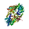

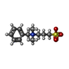


 Mass: 384.411 Da / Num. of mol.: 1 / Source method: obtained synthetically / Formula: C14H20N6O5S
 Mass: 257.349 Da / Num. of mol.: 1 / Source method: obtained synthetically / Formula: C12H19NO3S
 Mass: 120.147 Da / Num. of mol.: 1 / Source method: obtained synthetically / Formula: C5H12O3 / Comment: precipitant*YM
 Mass: 65.409 Da / Num. of mol.: 1 / Source method: obtained synthetically / Formula: Zn
 Sample preparation
 / Beamline: PROXIMA 1 / Wavelength: 0.979
  Processing