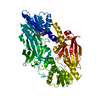
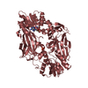
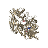
Deposited unit
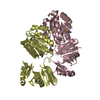
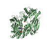
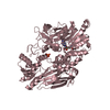
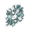
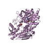
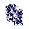
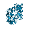
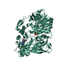
A: Protein arginine N-methyltransferase 7
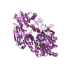
D: Protein arginine N-methyltransferase 7
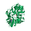
B: Protein arginine N-methyltransferase 7
C: Protein arginine N-methyltransferase 7
F: Protein arginine N-methyltransferase 7
E: Protein arginine N-methyltransferase 7
G: Protein arginine N-methyltransferase 7
H: Protein arginine N-methyltransferase 7
I: Protein arginine N-methyltransferase 7
M: Protein arginine N-methyltransferase 7
N: Protein arginine N-methyltransferase 7
O: Protein arginine N-methyltransferase 7
P: Protein arginine N-methyltransferase 7
Q: Protein arginine N-methyltransferase 7
R: Protein arginine N-methyltransferase 7
J: Protein arginine N-methyltransferase 7
K: Protein arginine N-methyltransferase 7
L: Protein arginine N-methyltransferase 7
hetero molecules Summary Component details
Theoretical mass Number of molelcules Total (without water) 1,342,338 54 Polymers 1,333,709 18 Non-polymers 8,629 36 Water 9,206 511
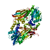
1
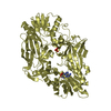
A: Protein arginine N-methyltransferase 7
hetero molecules Summary Component details Symmetry operations Calculated values
Theoretical mass Number of molelcules Total (without water) 74,574 3 Polymers 74,095 1 Non-polymers 479 2 Water 18 1
Type Name Symmetry operation Number identity operation 1_555 x,y,z 1
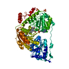
2
D: Protein arginine N-methyltransferase 7
hetero molecules Summary Component details Symmetry operations Calculated values
Theoretical mass Number of molelcules Total (without water) 74,574 3 Polymers 74,095 1 Non-polymers 479 2 Water 18 1
Type Name Symmetry operation Number identity operation 1_555 x,y,z 1
3
B: Protein arginine N-methyltransferase 7
hetero molecules Summary Component details Symmetry operations Calculated values
Theoretical mass Number of molelcules Total (without water) 74,574 3 Polymers 74,095 1 Non-polymers 479 2 Water 18 1
Type Name Symmetry operation Number identity operation 1_555 x,y,z 1
4
C: Protein arginine N-methyltransferase 7
hetero molecules Summary Component details Symmetry operations Calculated values
Theoretical mass Number of molelcules Total (without water) 74,574 3 Polymers 74,095 1 Non-polymers 479 2 Water 18 1
Type Name Symmetry operation Number identity operation 1_555 x,y,z 1
5
F: Protein arginine N-methyltransferase 7
hetero molecules Summary Component details Symmetry operations Calculated values
Theoretical mass Number of molelcules Total (without water) 74,574 3 Polymers 74,095 1 Non-polymers 479 2 Water 18 1
Type Name Symmetry operation Number identity operation 1_555 x,y,z 1
6
E: Protein arginine N-methyltransferase 7
hetero molecules Summary Component details Symmetry operations Calculated values
Theoretical mass Number of molelcules Total (without water) 74,574 3 Polymers 74,095 1 Non-polymers 479 2 Water 18 1
Type Name Symmetry operation Number identity operation 1_555 x,y,z 1
7
G: Protein arginine N-methyltransferase 7
hetero molecules Summary Component details Symmetry operations Calculated values
Theoretical mass Number of molelcules Total (without water) 74,574 3 Polymers 74,095 1 Non-polymers 479 2 Water 18 1
Type Name Symmetry operation Number identity operation 1_555 x,y,z 1
8
H: Protein arginine N-methyltransferase 7
hetero molecules Summary Component details Symmetry operations Calculated values
Theoretical mass Number of molelcules Total (without water) 74,574 3 Polymers 74,095 1 Non-polymers 479 2 Water 18 1
Type Name Symmetry operation Number identity operation 1_555 x,y,z 1
9
I: Protein arginine N-methyltransferase 7
hetero molecules Summary Component details Symmetry operations Calculated values
Theoretical mass Number of molelcules Total (without water) 74,574 3 Polymers 74,095 1 Non-polymers 479 2 Water 18 1
Type Name Symmetry operation Number identity operation 1_555 x,y,z 1
10
M: Protein arginine N-methyltransferase 7
hetero molecules Summary Component details Symmetry operations Calculated values
Theoretical mass Number of molelcules Total (without water) 74,574 3 Polymers 74,095 1 Non-polymers 479 2 Water 18 1
Type Name Symmetry operation Number identity operation 1_555 x,y,z 1
11
N: Protein arginine N-methyltransferase 7
hetero molecules Summary Component details Symmetry operations Calculated values
Theoretical mass Number of molelcules Total (without water) 74,574 3 Polymers 74,095 1 Non-polymers 479 2 Water 0
Type Name Symmetry operation Number identity operation 1_555 x,y,z 1
12
O: Protein arginine N-methyltransferase 7
hetero molecules Summary Component details Symmetry operations Calculated values
Theoretical mass Number of molelcules Total (without water) 74,574 3 Polymers 74,095 1 Non-polymers 479 2 Water 0
Type Name Symmetry operation Number identity operation 1_555 x,y,z 1
13
P: Protein arginine N-methyltransferase 7
hetero molecules Summary Component details Symmetry operations Calculated values
Theoretical mass Number of molelcules Total (without water) 74,574 3 Polymers 74,095 1 Non-polymers 479 2 Water 18 1
Type Name Symmetry operation Number identity operation 1_555 x,y,z 1
14
Q: Protein arginine N-methyltransferase 7
hetero molecules Summary Component details Symmetry operations Calculated values
Theoretical mass Number of molelcules Total (without water) 74,574 3 Polymers 74,095 1 Non-polymers 479 2 Water 18 1
Type Name Symmetry operation Number identity operation 1_555 x,y,z 1
15
R: Protein arginine N-methyltransferase 7
hetero molecules Summary Component details Symmetry operations Calculated values
Theoretical mass Number of molelcules Total (without water) 74,574 3 Polymers 74,095 1 Non-polymers 479 2 Water 18 1
Type Name Symmetry operation Number identity operation 1_555 x,y,z 1
16
J: Protein arginine N-methyltransferase 7
hetero molecules Summary Component details Symmetry operations Calculated values
Theoretical mass Number of molelcules Total (without water) 74,574 3 Polymers 74,095 1 Non-polymers 479 2 Water 18 1
Type Name Symmetry operation Number identity operation 1_555 x,y,z 1
17
K: Protein arginine N-methyltransferase 7
hetero molecules Summary Component details Symmetry operations Calculated values
Theoretical mass Number of molelcules Total (without water) 74,574 3 Polymers 74,095 1 Non-polymers 479 2 Water 18 1
Type Name Symmetry operation Number identity operation 1_555 x,y,z 1
18
L: Protein arginine N-methyltransferase 7
hetero molecules Summary Component details Symmetry operations Calculated values
Theoretical mass Number of molelcules Total (without water) 74,574 3 Polymers 74,095 1 Non-polymers 479 2 Water 18 1
Type Name Symmetry operation Number identity operation 1_555 x,y,z 1
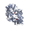
Unit cell Length a, b, c (Å) 190.703, 190.703, 373.088 Angle α, β, γ (deg.) 90.00, 90.00, 120.00 Int Tables number 144 Space group name H-M P31
 Movie
Movie Controller


 Open data
 Basic information
 Components
 Keywords
 Function and homology information
 X-RAY DIFFRACTION /
  Authors
 Citation
 Structure visualization
 Downloads & links
 Other downloads


 PDBj
PDBj Assembly
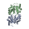
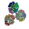


 Type: L-peptide linking / Mass: 384.411 Da / Num. of mol.: 18 / Source method: obtained synthetically / Formula: C14H20N6O5S
 Mass: 94.971 Da / Num. of mol.: 18 / Source method: obtained synthetically / Formula: PO4
 Mass: 18.015 Da / Num. of mol.: 511 / Source method: isolated from a natural source / Formula: H2O
 Sample preparation
 / Beamline: BL-5A / Wavelength: 1 Å
 Processing