 Movie
Movie Controller

[English] 日本語
 Yorodumi
- PDB-5xbj: The structure of the flagellar hook junction protein HAP1 (FlgK) ... -
+ Open data

- Basic information
| Entry | Database: PDB / ID: 5xbj | ||||||
|---|---|---|---|---|---|---|---|
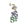
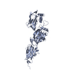
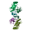
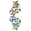
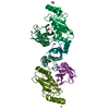
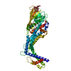
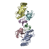
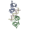
| Title | The structure of the flagellar hook junction protein HAP1 (FlgK) from Campylobacter jejuni | ||||||
 Components Components | Flagellar hook-associated protein FlgK | ||||||
 Keywords Keywords | BIOSYNTHETIC PROTEIN / Bacterial Flagellum / Hook / Filament / Junction protein | ||||||
| Function / homology |  Function and homology information Function and homology informationbacterial-type flagellum hook / bacterial-type flagellum assembly / structural molecule activity / extracellular region Similarity search - Function | ||||||
| Biological species |   Campylobacter jejuni (Campylobacter) Campylobacter jejuni (Campylobacter) | ||||||
| Method |  X-RAY DIFFRACTION / SYNCHROTRON / MOLECULAR REPLACEMENT / Resolution: 2.448 Å X-RAY DIFFRACTION / SYNCHROTRON / MOLECULAR REPLACEMENT / Resolution: 2.448 Å | ||||||
 Authors Authors | Samatey, F.A. | ||||||
| Funding support |  Japan, 1items Japan, 1items
| ||||||
 Citation Citation | Journal: Sci Rep / Year: 2017 Title: Structure of FlgK reveals the divergence of the bacterial Hook-Filament Junction of Campylobacter Authors: Bulieris, P.V. / Shaikh, N.H. / Freddolino, P.L. / Samatey, F.A. | ||||||
| History |
|
- Structure visualization
| Structure viewer | Molecule: MolmilJmol/JSmol |
|---|
- Downloads & links
-Download
| PDBx/mmCIF format | 5xbj.cif.gz | 121.9 KB | Display | PDBx/mmCIF format |
|---|---|---|---|---|
| PDB format | pdb5xbj.ent.gz | 91.4 KB | Display | PDB format |
| PDBx/mmJSON format | 5xbj.json.gz | Tree view | PDBx/mmJSON format | |
| Others |  Other downloads Other downloads |
-Validation report
| Arichive directory | https://data.pdbj.org/pub/pdb/validation_reports/xb/5xbjftp://data.pdbj.org/pub/pdb/validation_reports/xb/5xbj | HTTPS FTP |
|---|
-Related structure data
| Related structure data | 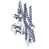 2d4yS S: Starting model for refinement |
|---|---|
| Similar structure data |
-Links
 PDBj
PDBj
- Assembly
| Deposited unit | 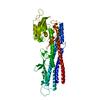
| |||||||||
|---|---|---|---|---|---|---|---|---|---|---|
| 1 |
| |||||||||
| Unit cell |
| |||||||||
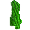
| Symmetry | Helical symmetry: (Circular symmetry: 1 / Dyad axis: no / N subunits divisor: 1 / Num. of operations: 11 / Rise per n subunits: 4.185 Å / Rotation per n subunits: 64.34 °) | |||||||||
| Components on special symmetry positions |
|
-Components
| #1: Protein | Mass: 56983.164 Da / Num. of mol.: 1 / Fragment: UNP RESIDUES 70-580 Source method: isolated from a genetically manipulated source Source: (gene. exp.) Campylobacter jejuni (Campylobacter)Production host: References: UniProt: A0A218KZD0, UniProt: Q0P8E9*PLUS |
|---|---|
| #2: Water | ChemComp-HOH /  Mass: 18.015 Da / Num. of mol.: 399 / Source method: isolated from a natural source / Formula: H2O Mass: 18.015 Da / Num. of mol.: 399 / Source method: isolated from a natural source / Formula: H2O |
-Experimental details
-Experiment
| Experiment | Method: X-RAY DIFFRACTION / Number of used crystals: 1 |
|---|
- Sample preparation
| Crystal | Density Matthews: 2.48 Å3/Da / Density % sol: 50.43 % |
|---|---|
| Crystal grow | Temperature: 293 K / Method: vapor diffusion, hanging drop / pH: 5.6 Details: 30% PEG MME 2000, 0.4M AMMONIUM, ACETATE, 2% MPD, 4% 2-PROPANOL, 5% ETHYLENE GLYCOL, 0.1M SODIUM CITRATE PH 5.6, VAPOR DIFFUSION, HANGING DROP, TEMPERATURE 293K |
-Data collection
| Diffraction | Mean temperature: 100 K |
|---|---|
| Diffraction source | Source: SYNCHROTRON / Site: SPring-8 / Beamline: BL44XU / Wavelength: 0.9 Å |
| Detector | Type: MARMOSAIC 300 mm CCD / Detector: CCD / Date: Mar 19, 2013 |
| Radiation | Protocol: SINGLE WAVELENGTH / Monochromatic (M) / Laue (L): M / Scattering type: x-ray |
| Radiation wavelength | Wavelength: 0.9 Å / Relative weight: 1 |
| Reflection | Resolution: 2.45→38.62 Å / Num. obs: 21573 / % possible obs: 99.76 % / Redundancy: 7.3 % / Net I/σ(I): 12.8 |
- Processing
| Software |
| |||||||||||||||||||||||||||||||||||||||||||||||||||||||||||||||||||||||||||||||||||||||||||||||||||||||||
|---|---|---|---|---|---|---|---|---|---|---|---|---|---|---|---|---|---|---|---|---|---|---|---|---|---|---|---|---|---|---|---|---|---|---|---|---|---|---|---|---|---|---|---|---|---|---|---|---|---|---|---|---|---|---|---|---|---|---|---|---|---|---|---|---|---|---|---|---|---|---|---|---|---|---|---|---|---|---|---|---|---|---|---|---|---|---|---|---|---|---|---|---|---|---|---|---|---|---|---|---|---|---|---|---|---|---|
| Refinement | Method to determine structure: MOLECULAR REPLACEMENT Starting model: 2D4Y Resolution: 2.448→19.971 Å / SU ML: 0.23 / Cross valid method: FREE R-VALUE / σ(F): 0 / Phase error: 20.77 / Stereochemistry target values: ML
| |||||||||||||||||||||||||||||||||||||||||||||||||||||||||||||||||||||||||||||||||||||||||||||||||||||||||
| Solvent computation | Shrinkage radii: 0.9 Å / VDW probe radii: 1.11 Å / Solvent model: FLAT BULK SOLVENT MODEL | |||||||||||||||||||||||||||||||||||||||||||||||||||||||||||||||||||||||||||||||||||||||||||||||||||||||||
| Refinement step | Cycle: LAST / Resolution: 2.448→19.971 Å
| |||||||||||||||||||||||||||||||||||||||||||||||||||||||||||||||||||||||||||||||||||||||||||||||||||||||||
| Refine LS restraints |
| |||||||||||||||||||||||||||||||||||||||||||||||||||||||||||||||||||||||||||||||||||||||||||||||||||||||||
| LS refinement shell |
|
