 Movie
Movie Controller

[English] 日本語
 Yorodumi
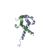
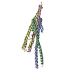
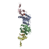
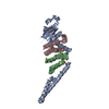
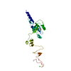
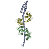
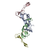
- PDB-6nkl: 2.2 A resolution structure of VapBC-1 from nontypeable Haemophilu... -
+ Open data

- Basic information
| Entry | Database: PDB / ID: 6nkl | |||||||||
|---|---|---|---|---|---|---|---|---|---|---|
| Title | 2.2 A resolution structure of VapBC-1 from nontypeable Haemophilus influenzae | |||||||||
 Components Components |
| |||||||||
 Keywords Keywords | ANTITOXIN / Toxin / H. influenzae / protein-protein complex | |||||||||
| Function / homology |  Function and homology information Function and homology informationRNA nuclease activity / toxin activity / Hydrolases; Acting on ester bonds / magnesium ion binding / DNA binding Similarity search - Function | |||||||||
| Biological species |  Haemophilus influenzae (bacteria) Haemophilus influenzae (bacteria) | |||||||||
| Method |  X-RAY DIFFRACTION / SYNCHROTRON / MOLECULAR REPLACEMENT / molecular replacement / Resolution: 2.2 Å X-RAY DIFFRACTION / SYNCHROTRON / MOLECULAR REPLACEMENT / molecular replacement / Resolution: 2.2 Å | |||||||||
 Authors Authors | Lovell, S. / Kashipathy, M.M. / Battaile, K.P. / Molinaro, A.L. / Daines, D.A. | |||||||||
| Funding support |  United States, 2items United States, 2items
| |||||||||
 Citation Citation | Journal: J.Bacteriol. / Year: 2019 Title: Crystal Structure of VapBC-1 from Nontypeable Haemophilus influenzae and the Effect of PIN Domain Mutations on Survival during Infection. Authors: Molinaro, A.L. / Kashipathy, M.M. / Lovell, S. / Battaile, K.P. / Coussens, N.P. / Shen, M. / Daines, D.A. | |||||||||
| History |
|
- Structure visualization
| Structure viewer | Molecule: MolmilJmol/JSmol |
|---|
- Downloads & links
-Download
| PDBx/mmCIF format | 6nkl.cif.gz | 96.4 KB | Display | PDBx/mmCIF format |
|---|---|---|---|---|
| PDB format | pdb6nkl.ent.gz | 70.7 KB | Display | PDB format |
| PDBx/mmJSON format | 6nkl.json.gz | Tree view | PDBx/mmJSON format | |
| Others |  Other downloads Other downloads |
-Validation report
| Arichive directory | https://data.pdbj.org/pub/pdb/validation_reports/nk/6nklftp://data.pdbj.org/pub/pdb/validation_reports/nk/6nkl | HTTPS FTP |
|---|
-Related structure data
| Related structure data |  5ecdS S: Starting model for refinement |
|---|---|
| Similar structure data |

-Links
 PDBj
PDBj

- Assembly
| Deposited unit | 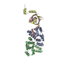
| ||||||||
|---|---|---|---|---|---|---|---|---|---|
| 1 | 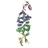
| ||||||||
| Unit cell |
|
-Components
| #1: Protein | Mass: 16894.568 Da / Num. of mol.: 2 / Fragment: VapB-1 Source method: isolated from a genetically manipulated source Details: Residues at the C-terminus (LLEHHHHHH) are from the purification tag. Source: (gene. exp.) Haemophilus influenzae (bacteria)Gene: vapC1, vapC, BV136_01367, BVZ80_01200, CH628_04345, NCTC11872_02278 Plasmid: pDD686 Details (production host): The VapB-1 and VapC-1 complex was coexpressed Production host: References: UniProt: A0A2R3FUY7, UniProt: Q57122*PLUS, Hydrolases; Acting on ester bonds #2: Protein | Mass: 10872.146 Da / Num. of mol.: 2 Source method: isolated from a genetically manipulated source Details: Residues (MASMTGG QQMGRDPNSS S) at the N-terminus are from the cloning vector. Source: (gene. exp.) Haemophilus influenzae (bacteria) / Gene: vapB1, BV136_01366, BVZ80_01199, CH628_04350 / Plasmid: pDD686Details (production host): The VapB-1 and VapC-1 complex was coexpressed Production host: #3: Water | ChemComp-HOH / |  Mass: 18.015 Da / Num. of mol.: 58 / Source method: isolated from a natural source / Formula: H2O Mass: 18.015 Da / Num. of mol.: 58 / Source method: isolated from a natural source / Formula: H2O |
|---|
-Experimental details
-Experiment
| Experiment | Method: X-RAY DIFFRACTION / Number of used crystals: 1 |
|---|
- Sample preparation
| Crystal | Density Matthews: 2 Å3/Da / Density % sol: 38.4 % |
|---|---|
| Crystal grow | Temperature: 291 K / Method: vapor diffusion, sitting drop / pH: 5 Details: 20% (w/v) PEG 4000, 0.1 M sodium acetate, 0.2 M ammonium acetate |
-Data collection
| Diffraction | Mean temperature: 100 K / Serial crystal experiment: N | |||||||||||||||||||||
|---|---|---|---|---|---|---|---|---|---|---|---|---|---|---|---|---|---|---|---|---|---|---|
| Diffraction source | Source: SYNCHROTRON / Site: APS / Beamline: 17-ID / Wavelength: 1 Å | |||||||||||||||||||||
| Detector | Type: DECTRIS PILATUS 6M / Detector: PIXEL / Date: Mar 4, 2018 | |||||||||||||||||||||
| Radiation | Protocol: SINGLE WAVELENGTH / Monochromatic (M) / Laue (L): M / Scattering type: x-ray | |||||||||||||||||||||
| Radiation wavelength | Wavelength: 1 Å / Relative weight: 1 | |||||||||||||||||||||
| Reflection | Resolution: 2.2→48.01 Å / Num. obs: 23388 / % possible obs: 100 % / Redundancy: 13 % / Biso Wilson estimate: 37.09 Å2 / CC1/2: 0.998 / Rmerge(I) obs: 0.168 / Net I/σ(I): 10.9 | |||||||||||||||||||||
| Reflection shell |
|
-Phasing
| Phasing | Method: molecular replacement | |||||||||
|---|---|---|---|---|---|---|---|---|---|---|
| Phasing MR | Model details: Phaser MODE: MR_AUTO
|
- Processing
| Software |
| ||||||||||||||||||||||||||||||||||||||||||||||||||||||
|---|---|---|---|---|---|---|---|---|---|---|---|---|---|---|---|---|---|---|---|---|---|---|---|---|---|---|---|---|---|---|---|---|---|---|---|---|---|---|---|---|---|---|---|---|---|---|---|---|---|---|---|---|---|---|---|
| Refinement | Method to determine structure: MOLECULAR REPLACEMENT Starting model: 5ECD Resolution: 2.2→48.01 Å / SU ML: 0.24 / Cross valid method: THROUGHOUT / σ(F): 1.01 / Phase error: 24.05
| ||||||||||||||||||||||||||||||||||||||||||||||||||||||
| Solvent computation | Shrinkage radii: 0.9 Å / VDW probe radii: 1.11 Å | ||||||||||||||||||||||||||||||||||||||||||||||||||||||
| Displacement parameters | Biso max: 95.71 Å2 / Biso mean: 43.4897 Å2 / Biso min: 20.7 Å2 | ||||||||||||||||||||||||||||||||||||||||||||||||||||||
| Refinement step | Cycle: final / Resolution: 2.2→48.01 Å
| ||||||||||||||||||||||||||||||||||||||||||||||||||||||
| LS refinement shell | Refine-ID: X-RAY DIFFRACTION / Rfactor Rfree error: 0 / Total num. of bins used: 8 / % reflection obs: 100 %
|
