 Movie
Movie Controller

[English] 日本語
 Yorodumi
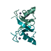
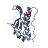
- PDB-7ai1: Crystal structure of human MDM2-G443T RING domain homodimer bound... -
+ Open data

- Basic information
| Entry | Database: PDB / ID: 7ai1 | ||||||||||||
|---|---|---|---|---|---|---|---|---|---|---|---|---|---|
| Title | Crystal structure of human MDM2-G443T RING domain homodimer bound to UbcH5B-Ub (Crystal form 2) | ||||||||||||
 Components Components |
| ||||||||||||
 Keywords Keywords | LIGASE / Ubiquitin ligase / RING E3 | ||||||||||||
| Function / homology |  Function and homology information Function and homology informationcellular response to vitamin B1 / response to formaldehyde / response to water-immersion restraint stress / response to ether / traversing start control point of mitotic cell cycle / atrial septum development / fibroblast activation / regulation of protein catabolic process at postsynapse, modulating synaptic transmission / Trafficking of AMPA receptors / (E3-independent) E2 ubiquitin-conjugating enzyme ...cellular response to vitamin B1 / response to formaldehyde / response to water-immersion restraint stress / response to ether / traversing start control point of mitotic cell cycle / atrial septum development / fibroblast activation / regulation of protein catabolic process at postsynapse, modulating synaptic transmission / Trafficking of AMPA receptors / (E3-independent) E2 ubiquitin-conjugating enzyme / receptor serine/threonine kinase binding / negative regulation of intrinsic apoptotic signaling pathway by p53 class mediator / negative regulation of protein processing / response to steroid hormone / SUMO transferase activity / peroxisome proliferator activated receptor binding / positive regulation of vascular associated smooth muscle cell migration / atrioventricular valve morphogenesis / AKT phosphorylates targets in the cytosol / hypothalamus gonadotrophin-releasing hormone neuron development / response to iron ion / NEDD8 ligase activity / female meiosis I / endocardial cushion morphogenesis / positive regulation of protein monoubiquitination / fat pad development / ventricular septum development / cellular response to peptide hormone stimulus / mitochondrion transport along microtubule / positive regulation of muscle cell differentiation / E2 ubiquitin-conjugating enzyme / regulation of postsynaptic neurotransmitter receptor internalization / cardiac septum morphogenesis / SUMOylation of ubiquitinylation proteins / seminiferous tubule development / cellular response to alkaloid / blood vessel development / ligase activity / Constitutive Signaling by AKT1 E17K in Cancer / female gonad development / negative regulation of DNA damage response, signal transduction by p53 class mediator / cellular response to antibiotic / SUMOylation of transcription factors / negative regulation of signal transduction by p53 class mediator / ubiquitin conjugating enzyme activity / regulation of protein catabolic process / cellular response to UV-C / male meiosis I / cellular response to estrogen stimulus / protein sumoylation / response to magnesium ion / blood vessel remodeling / positive regulation of intrinsic apoptotic signaling pathway by p53 class mediator / ribonucleoprotein complex binding / protein localization to nucleus / protein autoubiquitination / positive regulation of vascular associated smooth muscle cell proliferation / NPAS4 regulates expression of target genes / energy homeostasis / transcription repressor complex / neuron projection morphogenesis / protein K48-linked ubiquitination / regulation of proteasomal protein catabolic process / positive regulation of mitotic cell cycle / Maturation of protein E / Maturation of protein E / ER Quality Control Compartment (ERQC) / Myoclonic epilepsy of Lafora / FLT3 signaling by CBL mutants / IRAK2 mediated activation of TAK1 complex / regulation of heart rate / Alpha-protein kinase 1 signaling pathway / Glycogen synthesis / IRAK1 recruits IKK complex / IRAK1 recruits IKK complex upon TLR7/8 or 9 stimulation / Prevention of phagosomal-lysosomal fusion / Endosomal Sorting Complex Required For Transport (ESCRT) / Membrane binding and targetting of GAG proteins / Negative regulation of FLT3 / Regulation of TBK1, IKKε (IKBKE)-mediated activation of IRF3, IRF7 / PTK6 Regulates RTKs and Their Effectors AKT1 and DOK1 / Regulation of TBK1, IKKε-mediated activation of IRF3, IRF7 upon TLR3 ligation / IRAK2 mediated activation of TAK1 complex upon TLR7/8 or 9 stimulation / Constitutive Signaling by NOTCH1 HD Domain Mutants / NOTCH2 Activation and Transmission of Signal to the Nucleus / TICAM1,TRAF6-dependent induction of TAK1 complex / : / TICAM1-dependent activation of IRF3/IRF7 / APC/C:Cdc20 mediated degradation of Cyclin B / regulation of neuron apoptotic process / Downregulation of ERBB4 signaling / APC-Cdc20 mediated degradation of Nek2A / Regulation of FZD by ubiquitination / positive regulation of protein export from nucleus / p75NTR recruits signalling complexes / InlA-mediated entry of Listeria monocytogenes into host cells / TRAF6 mediated IRF7 activation in TLR7/8 or 9 signaling / NF-kB is activated and signals survival / TRAF6-mediated induction of TAK1 complex within TLR4 complex / Regulation of pyruvate metabolism Similarity search - Function | ||||||||||||
| Biological species |  Homo sapiens (human) Homo sapiens (human) | ||||||||||||
| Method |  X-RAY DIFFRACTION / SYNCHROTRON / MOLECULAR REPLACEMENT / Resolution: 2.07 Å X-RAY DIFFRACTION / SYNCHROTRON / MOLECULAR REPLACEMENT / Resolution: 2.07 Å | ||||||||||||
 Authors Authors | Magnussen, H.M. / Huang, D.T. | ||||||||||||
| Funding support |  United Kingdom, 3items United Kingdom, 3items
| ||||||||||||
 Citation Citation | Journal: J.Mol.Biol. / Year: 2021 Title: Identification of a Catalytic Active but Non-Aggregating MDM2 RING Domain Variant. Authors: Magnussen, H.M. / Huang, D.T. | ||||||||||||
| History |
|
- Structure visualization
| Structure viewer | Molecule: MolmilJmol/JSmol |
|---|
- Downloads & links
-Download
| PDBx/mmCIF format | 7ai1.cif.gz | 240.6 KB | Display | PDBx/mmCIF format |
|---|---|---|---|---|
| PDB format | pdb7ai1.ent.gz | Display | PDB format | |
| PDBx/mmJSON format | 7ai1.json.gz | Tree view | PDBx/mmJSON format | |
| Others |  Other downloads Other downloads |
-Validation report
| Arichive directory | https://data.pdbj.org/pub/pdb/validation_reports/ai/7ai1ftp://data.pdbj.org/pub/pdb/validation_reports/ai/7ai1 | HTTPS FTP |
|---|
-Related structure data


-Links
 PDBj
PDBj


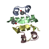
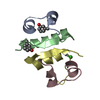
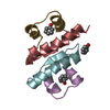
- Assembly
| Deposited unit | 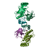
| ||||||||||||||||
|---|---|---|---|---|---|---|---|---|---|---|---|---|---|---|---|---|---|
| 1 | 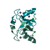
| ||||||||||||||||
| 2 | 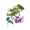
| ||||||||||||||||
| 3 | 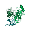
| ||||||||||||||||
| Unit cell |
| ||||||||||||||||
| Noncrystallographic symmetry (NCS) | NCS domain:
NCS ensembles :
|
-Components
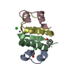
-Protein , 3 types, 6 molecules AAADDDBBBEEECCCFFF
| #1: Protein | Mass: 8329.913 Da / Num. of mol.: 2 / Mutation: G443T Source method: isolated from a genetically manipulated source Details: G417, S418: Expression tag / Source: (gene. exp.) Homo sapiens (human) / Gene: MDM2 / Production host:  References: UniProt: Q00987, RING-type E3 ubiquitin transferase #2: Protein | Mass: 16851.381 Da / Num. of mol.: 2 / Mutation: S22R, C85K Source method: isolated from a genetically manipulated source Details: C85K is covalently linked to G76 (Molecule 3). / Source: (gene. exp.) Homo sapiens (human) / Gene: UBE2D2, PUBC1, UBC4, UBC5B, UBCH4, UBCH5B / Production host: References: UniProt: P62837, E2 ubiquitin-conjugating enzyme, (E3-independent) E2 ubiquitin-conjugating enzyme #3: Protein | Mass: 8922.141 Da / Num. of mol.: 2 Source method: isolated from a genetically manipulated source Details: G-4,S-3,G-2,G-1,S0: Expression tag G76 is covalently linked to C85K (Molecule 2). Source: (gene. exp.) Homo sapiens (human) / Gene: UBB / Production host: |
|---|
-Non-polymers , 4 types, 286 molecules 


| #4: Chemical |  Mass: 35.453 Da / Num. of mol.: 2 / Source method: obtained synthetically / Formula: Cl Mass: 35.453 Da / Num. of mol.: 2 / Source method: obtained synthetically / Formula: Cl#5: Chemical | ChemComp-ZN /  Mass: 65.409 Da / Num. of mol.: 4 / Source method: obtained synthetically / Formula: Zn Mass: 65.409 Da / Num. of mol.: 4 / Source method: obtained synthetically / Formula: Zn#6: Chemical |  Mass: 62.068 Da / Num. of mol.: 2 / Source method: obtained synthetically / Formula: C2H6O2 Mass: 62.068 Da / Num. of mol.: 2 / Source method: obtained synthetically / Formula: C2H6O2#7: Water | ChemComp-HOH / | Mass: 18.015 Da / Num. of mol.: 278 / Source method: isolated from a natural source / Formula: H2O |
|---|
-Details
| Has ligand of interest | N |
|---|
-Experimental details
-Experiment
| Experiment | Method: X-RAY DIFFRACTION / Number of used crystals: 1 |
|---|
- Sample preparation
| Crystal | Density Matthews: 2.26 Å3/Da / Density % sol: 45.61 % |
|---|---|
| Crystal grow | Temperature: 292 K / Method: vapor diffusion, sitting drop / pH: 7.5 Details: 0.1 M HEPES, 0.2 M NH4NO3, 20 % w/v PEG Smear Broad |
-Data collection
| Diffraction | Mean temperature: 100 K / Serial crystal experiment: N |
|---|---|
| Diffraction source | Source: SYNCHROTRON / Site: Diamond / Beamline: I04-1 / Wavelength: 0.91587 Å |
| Detector | Type: DECTRIS PILATUS 6M-F / Detector: PIXEL / Date: Mar 9, 2018 |
| Radiation | Protocol: SINGLE WAVELENGTH / Monochromatic (M) / Laue (L): M / Scattering type: x-ray |
| Radiation wavelength | Wavelength: 0.91587 Å / Relative weight: 1 |
| Reflection | Resolution: 2.07→135.91 Å / Num. obs: 38548 / % possible obs: 100 % / Redundancy: 6.4 % / CC1/2: 0.997 / Net I/σ(I): 9 |
| Reflection shell | Resolution: 2.07→2.11 Å / Num. unique obs: 1861 / CC1/2: 0.584 |
- Processing
| Software |
| |||||||||||||||||||||||||||||||||||||||||||||||||||||||||||||||||||||||||||||||||||||||||||||||||||||||||||||||||||||||||||||||||||||||||||||||||||||||||||||||||||||||||||||||
|---|---|---|---|---|---|---|---|---|---|---|---|---|---|---|---|---|---|---|---|---|---|---|---|---|---|---|---|---|---|---|---|---|---|---|---|---|---|---|---|---|---|---|---|---|---|---|---|---|---|---|---|---|---|---|---|---|---|---|---|---|---|---|---|---|---|---|---|---|---|---|---|---|---|---|---|---|---|---|---|---|---|---|---|---|---|---|---|---|---|---|---|---|---|---|---|---|---|---|---|---|---|---|---|---|---|---|---|---|---|---|---|---|---|---|---|---|---|---|---|---|---|---|---|---|---|---|---|---|---|---|---|---|---|---|---|---|---|---|---|---|---|---|---|---|---|---|---|---|---|---|---|---|---|---|---|---|---|---|---|---|---|---|---|---|---|---|---|---|---|---|---|---|---|---|---|---|
| Refinement | Method to determine structure: MOLECULAR REPLACEMENT Starting model: MDM2-E2-Ub Resolution: 2.07→69.478 Å / Cor.coef. Fo:Fc: 0.947 / Cor.coef. Fo:Fc free: 0.921 / WRfactor Rfree: 0.241 / WRfactor Rwork: 0.191 / SU B: 6.44 / SU ML: 0.165 / Average fsc free: 0.8728 / Average fsc work: 0.8939 / Cross valid method: FREE R-VALUE / ESU R: 0.234 / ESU R Free: 0.203 Details: Hydrogens have been added in their riding positions
| |||||||||||||||||||||||||||||||||||||||||||||||||||||||||||||||||||||||||||||||||||||||||||||||||||||||||||||||||||||||||||||||||||||||||||||||||||||||||||||||||||||||||||||||
| Solvent computation | Ion probe radii: 0.8 Å / Shrinkage radii: 0.8 Å / VDW probe radii: 1.2 Å / Solvent model: MASK BULK SOLVENT | |||||||||||||||||||||||||||||||||||||||||||||||||||||||||||||||||||||||||||||||||||||||||||||||||||||||||||||||||||||||||||||||||||||||||||||||||||||||||||||||||||||||||||||||
| Displacement parameters | Biso mean: 32.5 Å2
| |||||||||||||||||||||||||||||||||||||||||||||||||||||||||||||||||||||||||||||||||||||||||||||||||||||||||||||||||||||||||||||||||||||||||||||||||||||||||||||||||||||||||||||||
| Refinement step | Cycle: LAST / Resolution: 2.07→69.478 Å
| |||||||||||||||||||||||||||||||||||||||||||||||||||||||||||||||||||||||||||||||||||||||||||||||||||||||||||||||||||||||||||||||||||||||||||||||||||||||||||||||||||||||||||||||
| Refine LS restraints |
| |||||||||||||||||||||||||||||||||||||||||||||||||||||||||||||||||||||||||||||||||||||||||||||||||||||||||||||||||||||||||||||||||||||||||||||||||||||||||||||||||||||||||||||||
| LS refinement shell |
|
