 Movie
Movie Controller

[English] 日本語
 Yorodumi
- PDB-5xax: Parallel homodimer structures of the extracellular domains of the... -
+ Open data

- Basic information
| Entry | Database: PDB / ID: 5xax | ||||||
|---|---|---|---|---|---|---|---|
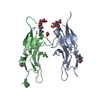
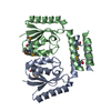
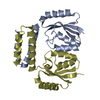
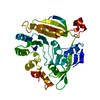
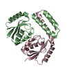
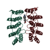
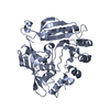
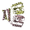
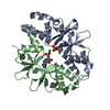
| Title | Parallel homodimer structures of the extracellular domains of the voltage-gated sodium channel beta4 subunit explain its role in cell-cell adhesion | ||||||
 Components Components | Sodium channel subunit beta-4 | ||||||
 Keywords Keywords | CELL ADHESION / Ig | ||||||
| Function / homology |  Function and homology information Function and homology informationAV node cell action potential / voltage-gated sodium channel activity involved in cardiac muscle cell action potential / cardiac conduction / membrane depolarization during cardiac muscle cell action potential / positive regulation of sodium ion transport / regulation of ventricular cardiac muscle cell membrane repolarization / cardiac muscle cell action potential involved in contraction / voltage-gated sodium channel complex / voltage-gated sodium channel activity / sodium ion transport ...AV node cell action potential / voltage-gated sodium channel activity involved in cardiac muscle cell action potential / cardiac conduction / membrane depolarization during cardiac muscle cell action potential / positive regulation of sodium ion transport / regulation of ventricular cardiac muscle cell membrane repolarization / cardiac muscle cell action potential involved in contraction / voltage-gated sodium channel complex / voltage-gated sodium channel activity / sodium ion transport / regulation of heart rate by cardiac conduction / intercalated disc / sodium channel regulator activity / neuronal action potential / cardiac muscle contraction / sodium ion transmembrane transport / transmembrane transporter binding / plasma membrane Similarity search - Function | ||||||
| Biological species |  | ||||||
| Method |  X-RAY DIFFRACTION / SYNCHROTRON / MOLECULAR REPLACEMENT / Resolution: 2.903 Å X-RAY DIFFRACTION / SYNCHROTRON / MOLECULAR REPLACEMENT / Resolution: 2.903 Å | ||||||
 Authors Authors | Shimizu, H. / Yokoyama, S. | ||||||
 Citation Citation | Journal: J. Biol. Chem. / Year: 2017 Title: Parallel homodimer structures of the extracellular domains of the voltage-gated sodium channel beta 4 subunit explain its role in cell-cell adhesion Authors: Shimizu, H. / Tosaki, A. / Ohsawa, N. / Ishizuka-Katsura, Y. / Shoji, S. / Miyazaki, H. / Oyama, F. / Terada, T. / Shirouzu, M. / Sekine, S.I. / Nukina, N. / Yokoyama, S. | ||||||
| History |
|
- Structure visualization
| Structure viewer | Molecule: MolmilJmol/JSmol |
|---|
- Downloads & links
-Download
| PDBx/mmCIF format | 5xax.cif.gz | 60.8 KB | Display | PDBx/mmCIF format |
|---|---|---|---|---|
| PDB format | pdb5xax.ent.gz | 44.5 KB | Display | PDB format |
| PDBx/mmJSON format | 5xax.json.gz | Tree view | PDBx/mmJSON format | |
| Others |  Other downloads Other downloads |
-Validation report
| Arichive directory | https://data.pdbj.org/pub/pdb/validation_reports/xa/5xaxftp://data.pdbj.org/pub/pdb/validation_reports/xa/5xax | HTTPS FTP |
|---|
-Related structure data
| Related structure data |  5xawC  5ayqS C: citing same article ( S: Starting model for refinement |
|---|---|
| Similar structure data |

-Links
 PDBj
PDBj
- Assembly
| Deposited unit | 
| ||||||||
|---|---|---|---|---|---|---|---|---|---|
| 1 |
| ||||||||
| Unit cell |
|
-Components
| #1: Protein | Mass: 15791.683 Da / Num. of mol.: 2 / Fragment: Extracellula domain, UNP residues 30-160 Source method: isolated from a genetically manipulated source Source: (gene. exp.) Production host:  Strain (production host): BL21-Gold(DE3)pLysS AG / References: UniProt: Q7M729 #2: Chemical | ChemComp-GOL / |   Mass: 92.094 Da / Num. of mol.: 1 / Source method: obtained synthetically / Formula: C3H8O3 Mass: 92.094 Da / Num. of mol.: 1 / Source method: obtained synthetically / Formula: C3H8O3Has protein modification | Y | |
|---|
-Experimental details
-Experiment
| Experiment | Method: X-RAY DIFFRACTION / Number of used crystals: 1 |
|---|
- Sample preparation
| Crystal | Density Matthews: 3 Å3/Da / Density % sol: 59.04 % |
|---|---|
| Crystal grow | Temperature: 293 K / Method: vapor diffusion, sitting drop / pH: 7 Details: 2.7M Na-malonate, 0.1M Tris-HCl (pH 7.0), 0.5 % (v/v) Tween 20 |
-Data collection
| Diffraction | Mean temperature: 100 K |
|---|---|
| Diffraction source | Source: SYNCHROTRON / Site: SPring-8  / Beamline: BL44B2 / Wavelength: 0.8 Å / Beamline: BL44B2 / Wavelength: 0.8 Å |
| Detector | Type: MAR CCD 165 mm / Detector: CCD / Date: Dec 22, 2004 |
| Radiation | Protocol: SINGLE WAVELENGTH / Monochromatic (M) / Laue (L): M / Scattering type: x-ray |
| Radiation wavelength | Wavelength: 0.8 Å / Relative weight: 1 |
| Reflection | Resolution: 2.9→35.2 Å / Num. obs: 8526 / % possible obs: 99.6 % / Redundancy: 7.8 % / Net I/σ(I): 31.4 |
- Processing
| Software |
| |||||||||||||||||||||||||||||||||||||||||||||||||
|---|---|---|---|---|---|---|---|---|---|---|---|---|---|---|---|---|---|---|---|---|---|---|---|---|---|---|---|---|---|---|---|---|---|---|---|---|---|---|---|---|---|---|---|---|---|---|---|---|---|---|
| Refinement | Method to determine structure: MOLECULAR REPLACEMENT Starting model: 5AYQ Resolution: 2.903→35.157 Å / SU ML: 0.46 / Cross valid method: FREE R-VALUE / σ(F): 1.34 / Phase error: 32.35 / Stereochemistry target values: ML
| |||||||||||||||||||||||||||||||||||||||||||||||||
| Solvent computation | Shrinkage radii: 0.9 Å / VDW probe radii: 1.11 Å / Solvent model: FLAT BULK SOLVENT MODEL | |||||||||||||||||||||||||||||||||||||||||||||||||
| Refinement step | Cycle: LAST / Resolution: 2.903→35.157 Å
| |||||||||||||||||||||||||||||||||||||||||||||||||
| Refine LS restraints |
| |||||||||||||||||||||||||||||||||||||||||||||||||
| LS refinement shell |
|
