 Movie
Movie Controller

[English] 日本語
 Yorodumi
- PDB-5x9i: Unique Choloylglycine Hydrolase(CGH) member Mutant (C1S) from She... -
+ Open data

- Basic information
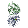
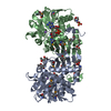
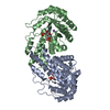
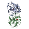
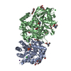
| Entry | Database: PDB / ID: 5x9i | ||||||
|---|---|---|---|---|---|---|---|
| Title | Unique Choloylglycine Hydrolase(CGH) member Mutant (C1S) from Shewanella loihica PV-4 | ||||||
 Components Components | Penicillin V acylase-like protein | ||||||
 Keywords Keywords | HYDROLASE / Choloylglycine Hydrolase / BSH / PVA like / AHL activity / C1S mutant | ||||||
| Function / homology | : / Choloylglycine hydrolase/NAAA C-terminal / Linear amide C-N hydrolases, choloylglycine hydrolase family / Nucleophile aminohydrolases, N-terminal / hydrolase activity / 3-OXOOCTANOIC ACID / Penicillin V acylase-like protein Function and homology information Function and homology information | ||||||
| Biological species |  Shewanella loihica PV-4 (bacteria) Shewanella loihica PV-4 (bacteria) | ||||||
| Method |  X-RAY DIFFRACTION / SYNCHROTRON / MOLECULAR REPLACEMENT / Resolution: 1.5 Å X-RAY DIFFRACTION / SYNCHROTRON / MOLECULAR REPLACEMENT / Resolution: 1.5 Å | ||||||
 Authors Authors | Ramasamy, S. / Philem, P. / Yadav, Y. | ||||||
 Citation Citation | Journal: To Be Published Title: Unique Choloylglycine Hydrolase(CGH) member from Shewanella loihica PV-4 Authors: Philem, P. / Yadav, Y. / Prabune, A.A. / Ramasamy, S. | ||||||
| History |
|
- Structure visualization
| Structure viewer | Molecule: MolmilJmol/JSmol |
|---|
- Downloads & links
-Download
| PDBx/mmCIF format | 5x9i.cif.gz | 145.8 KB | Display | PDBx/mmCIF format |
|---|---|---|---|---|
| PDB format | pdb5x9i.ent.gz | 113.7 KB | Display | PDB format |
| PDBx/mmJSON format | 5x9i.json.gz | Tree view | PDBx/mmJSON format | |
| Others |  Other downloads Other downloads |
-Validation report
| Arichive directory | https://data.pdbj.org/pub/pdb/validation_reports/x9/5x9iftp://data.pdbj.org/pub/pdb/validation_reports/x9/5x9i | HTTPS FTP |
|---|
-Related structure data
| Related structure data |  5x8zC  3hbcS S: Starting model for refinement C: citing same article ( |
|---|---|
| Similar structure data |


-Links
 PDBj
PDBj


- Assembly
| Deposited unit | 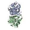
| ||||||||
|---|---|---|---|---|---|---|---|---|---|
| 1 |
| ||||||||
| Unit cell |
|
-Components
| #1: Protein | Mass: 37928.387 Da / Num. of mol.: 2 / Fragment: UNP residues 27-359 / Mutation: C1S Source method: isolated from a genetically manipulated source Source: (gene. exp.) Shewanella loihica PV-4 (bacteria) / Strain: PV-4 / Gene: Shew_0681 / Plasmid: pET22b+ / Production host: #2: Chemical | 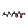  Mass: 158.195 Da / Num. of mol.: 2 / Source method: obtained synthetically / Formula: C8H14O3 Mass: 158.195 Da / Num. of mol.: 2 / Source method: obtained synthetically / Formula: C8H14O3#3: Chemical |   Mass: 92.094 Da / Num. of mol.: 2 / Source method: isolated from a natural source / Formula: C3H8O3 Mass: 92.094 Da / Num. of mol.: 2 / Source method: isolated from a natural source / Formula: C3H8O3#4: Water | ChemComp-HOH / |  Mass: 18.015 Da / Num. of mol.: 175 / Source method: isolated from a natural source / Formula: H2O Mass: 18.015 Da / Num. of mol.: 175 / Source method: isolated from a natural source / Formula: H2O |
|---|
-Experimental details
-Experiment
| Experiment | Method: X-RAY DIFFRACTION / Number of used crystals: 1 |
|---|
- Sample preparation
| Crystal | Density Matthews: 2.38 Å3/Da / Density % sol: 48.39 % / Description: elongated needle crystal |
|---|---|
| Crystal grow | Temperature: 293.15 K / Method: vapor diffusion, hanging drop / pH: 7.2 Details: PEG 3350, sodium sulphate, 2-Methyl-2, 4-pentanediol |
-Data collection
| Diffraction | Mean temperature: 100 K |
|---|---|
| Diffraction source | Source: SYNCHROTRON / Site: ESRF  / Beamline: MASSIF-3 / Wavelength: 0.97855 Å / Beamline: MASSIF-3 / Wavelength: 0.97855 Å |
| Detector | Type: RAYONIX MX-225 / Detector: FLAT PANEL / Date: Feb 28, 2016 |
| Radiation | Monochromator: Si(111) single crystal monochromator / Protocol: SINGLE WAVELENGTH / Monochromatic (M) / Laue (L): M / Scattering type: x-ray |
| Radiation wavelength | Wavelength: 0.97855 Å / Relative weight: 1 |
| Reflection | Resolution: 1.5→50 Å / Num. obs: 105996 / % possible obs: 100 % / Redundancy: 4.9 % / Net I/σ(I): 12.2 |
- Processing
| Software |
| ||||||||||||||||||||||||||||||||||||||||||||||||||||||||||||||||||||||||||||||||||||||||||||||||||||||||||||||||||||||||||||||||||||||||||||||||||||||||||||||||||||||||||||||||||||||
|---|---|---|---|---|---|---|---|---|---|---|---|---|---|---|---|---|---|---|---|---|---|---|---|---|---|---|---|---|---|---|---|---|---|---|---|---|---|---|---|---|---|---|---|---|---|---|---|---|---|---|---|---|---|---|---|---|---|---|---|---|---|---|---|---|---|---|---|---|---|---|---|---|---|---|---|---|---|---|---|---|---|---|---|---|---|---|---|---|---|---|---|---|---|---|---|---|---|---|---|---|---|---|---|---|---|---|---|---|---|---|---|---|---|---|---|---|---|---|---|---|---|---|---|---|---|---|---|---|---|---|---|---|---|---|---|---|---|---|---|---|---|---|---|---|---|---|---|---|---|---|---|---|---|---|---|---|---|---|---|---|---|---|---|---|---|---|---|---|---|---|---|---|---|---|---|---|---|---|---|---|---|---|---|
| Refinement | Method to determine structure: MOLECULAR REPLACEMENT Starting model: 3HBC Resolution: 1.5→35.891 Å / Cor.coef. Fo:Fc: 0.966 / Cor.coef. Fo:Fc free: 0.961 / Cross valid method: THROUGHOUT / ESU R: 0.072 / ESU R Free: 0.072 / Stereochemistry target values: MAXIMUM LIKELIHOOD / Details: HYDROGENS HAVE BEEN ADDED IN THE RIDING POSITIONS
| ||||||||||||||||||||||||||||||||||||||||||||||||||||||||||||||||||||||||||||||||||||||||||||||||||||||||||||||||||||||||||||||||||||||||||||||||||||||||||||||||||||||||||||||||||||||
| Solvent computation | Ion probe radii: 0.8 Å / Shrinkage radii: 0.8 Å / VDW probe radii: 1.2 Å / Solvent model: MASK | ||||||||||||||||||||||||||||||||||||||||||||||||||||||||||||||||||||||||||||||||||||||||||||||||||||||||||||||||||||||||||||||||||||||||||||||||||||||||||||||||||||||||||||||||||||||
| Displacement parameters | Biso mean: 19.563 Å2
| ||||||||||||||||||||||||||||||||||||||||||||||||||||||||||||||||||||||||||||||||||||||||||||||||||||||||||||||||||||||||||||||||||||||||||||||||||||||||||||||||||||||||||||||||||||||
| Refinement step | Cycle: 1 / Resolution: 1.5→35.891 Å
| ||||||||||||||||||||||||||||||||||||||||||||||||||||||||||||||||||||||||||||||||||||||||||||||||||||||||||||||||||||||||||||||||||||||||||||||||||||||||||||||||||||||||||||||||||||||
| Refine LS restraints |
|