 Movie
Movie Controller


+ Open data

- Basic information
| Entry | Database: PDB / ID: 1wgj | ||||||
|---|---|---|---|---|---|---|---|
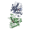
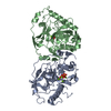
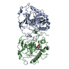
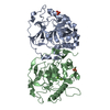
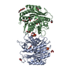
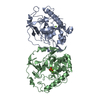
| Title | STRUCTURE OF INORGANIC PYROPHOSPHATASE | ||||||
 Components Components | INORGANIC PYROPHOSPHATASE | ||||||
 Keywords Keywords | HYDROLASE / PYROPHOSPHATE PHOSPHOHYDROLASE / MANGANESE | ||||||
| Function / homology |  Function and homology information Function and homology informationCytosolic tRNA aminoacylation / Mitochondrial tRNA aminoacylation / Pyrophosphate hydrolysis / inorganic diphosphatase / inorganic diphosphate phosphatase activity / phosphate-containing compound metabolic process / magnesium ion binding / nucleus / cytoplasm Similarity search - Function | ||||||
| Biological species |  | ||||||
| Method |  X-RAY DIFFRACTION / Resolution: 2 Å X-RAY DIFFRACTION / Resolution: 2 Å | ||||||
 Authors Authors | Heikinheimo, P. / Lehtonen, J. / Goldman, A. | ||||||
 Citation Citation | Journal: Structure / Year: 1996 Title: The structural basis for pyrophosphatase catalysis. Authors: Heikinheimo, P. / Lehtonen, J. / Baykov, A. / Lahti, R. / Cooperman, B.S. / Goldman, A. #1: Journal: Acta Crystallogr.,Sect.D / Year: 1995Title: New Crystal Forms of E. Coli and S. Cerevisiae Soluble Inorganic Pyrophosphatases Authors: Heikinheimo, P. / Salminen, T. / Cooperman, B.S. / Lahti, R. / Goldman, A. | ||||||
| History |
|
- Structure visualization
| Structure viewer | Molecule: MolmilJmol/JSmol |
|---|
- Downloads & links
-Download
| PDBx/mmCIF format | 1wgj.cif.gz | 129.6 KB | Display | PDBx/mmCIF format |
|---|---|---|---|---|
| PDB format | pdb1wgj.ent.gz | 101.2 KB | Display | PDB format |
| PDBx/mmJSON format | 1wgj.json.gz | Tree view | PDBx/mmJSON format | |
| Others |  Other downloads Other downloads |
-Validation report
| Arichive directory | https://data.pdbj.org/pub/pdb/validation_reports/wg/1wgjftp://data.pdbj.org/pub/pdb/validation_reports/wg/1wgj | HTTPS FTP |
|---|
-Related structure data


-Links
 PDBj
PDBj- Assembly
| Deposited unit | 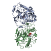
| ||||||||
|---|---|---|---|---|---|---|---|---|---|
| 1 |
| ||||||||
| Unit cell |
|
-Components
| #1: Protein | Mass: 32225.375 Da / Num. of mol.: 2 Source method: isolated from a genetically manipulated source Details: PRODUCT COMPLEX Source: (gene. exp.) Gene: PPA1 / Plasmid: PKW9 / Gene (production host): PPA1 / Production host:  #2: Chemical | ChemComp-MN /   Mass: 54.938 Da / Num. of mol.: 8 / Source method: obtained synthetically / Formula: Mn Mass: 54.938 Da / Num. of mol.: 8 / Source method: obtained synthetically / Formula: Mn#3: Chemical | ChemComp-PO4 /   Mass: 94.971 Da / Num. of mol.: 4 / Source method: obtained synthetically / Formula: PO4 Mass: 94.971 Da / Num. of mol.: 4 / Source method: obtained synthetically / Formula: PO4#4: Water | ChemComp-HOH / |  Mass: 18.015 Da / Num. of mol.: 307 / Source method: isolated from a natural source / Formula: H2O Mass: 18.015 Da / Num. of mol.: 307 / Source method: isolated from a natural source / Formula: H2O |
|---|
-Experimental details
-Experiment
| Experiment | Method: X-RAY DIFFRACTION |
|---|
- Sample preparation
| Crystal | Density Matthews: 2.79 Å3/Da / Density % sol: 55.87 % | ||||||||||||||||||||||||||||||||||||
|---|---|---|---|---|---|---|---|---|---|---|---|---|---|---|---|---|---|---|---|---|---|---|---|---|---|---|---|---|---|---|---|---|---|---|---|---|---|
| Crystal grow | *PLUS pH: 6 / Method: vapor diffusion, sitting drop | ||||||||||||||||||||||||||||||||||||
| Components of the solutions | *PLUS
|
-Data collection
| Diffraction source | Wavelength: 1.5418 |
|---|---|
| Detector | Type: RIGAKU / Detector: IMAGE PLATE |
| Radiation | Monochromatic (M) / Laue (L): M / Scattering type: x-ray |
| Radiation wavelength | Wavelength: 1.5418 Å / Relative weight: 1 |
| Reflection | Redundancy: 5.2 % / Biso Wilson estimate: 5.8 Å2 / Rmerge(I) obs: 0.103 |
| Reflection | *PLUS Highest resolution: 2 Å / Lowest resolution: 8 Å / Num. obs: 45565 / % possible obs: 89.2 % / Num. measured all: 236904 |
| Reflection shell | *PLUS Highest resolution: 2 Å / Lowest resolution: 2.09 Å / % possible obs: 82.9 % |
- Processing
| Software |
| ||||||||||||||||||||||||||||||||||||||||||||||||||||||||||||||||||||||||||||||||
|---|---|---|---|---|---|---|---|---|---|---|---|---|---|---|---|---|---|---|---|---|---|---|---|---|---|---|---|---|---|---|---|---|---|---|---|---|---|---|---|---|---|---|---|---|---|---|---|---|---|---|---|---|---|---|---|---|---|---|---|---|---|---|---|---|---|---|---|---|---|---|---|---|---|---|---|---|---|---|---|---|---|
| Refinement | Resolution: 2→8 Å / Rfactor Rfree error: 0.005 / Data cutoff high absF: 100000 / Data cutoff low absF: 0.1 / Isotropic thermal model: RESTRAINED / Cross valid method: THROUGHOUT / σ(F): 2
| ||||||||||||||||||||||||||||||||||||||||||||||||||||||||||||||||||||||||||||||||
| Displacement parameters | Biso mean: 18.1 Å2
| ||||||||||||||||||||||||||||||||||||||||||||||||||||||||||||||||||||||||||||||||
| Refine analyze |
| ||||||||||||||||||||||||||||||||||||||||||||||||||||||||||||||||||||||||||||||||
| Refinement step | Cycle: LAST / Resolution: 2→8 Å
| ||||||||||||||||||||||||||||||||||||||||||||||||||||||||||||||||||||||||||||||||
| Refine LS restraints |
| ||||||||||||||||||||||||||||||||||||||||||||||||||||||||||||||||||||||||||||||||
| LS refinement shell | Resolution: 2→2.09 Å / Rfactor Rfree error: 0.015 / Total num. of bins used: 8
| ||||||||||||||||||||||||||||||||||||||||||||||||||||||||||||||||||||||||||||||||
| Xplor file |
| ||||||||||||||||||||||||||||||||||||||||||||||||||||||||||||||||||||||||||||||||
| Software | *PLUS Name: X-PLOR / Version: 3.1 / Classification: refinement | ||||||||||||||||||||||||||||||||||||||||||||||||||||||||||||||||||||||||||||||||
| Refine LS restraints | *PLUS
|
