 Movie
Movie Controller


+ Open data

- Basic information
| Entry | Database: PDB / ID: 1e9g | ||||||
|---|---|---|---|---|---|---|---|
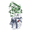
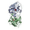
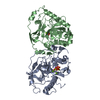
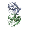
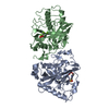
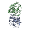
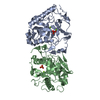
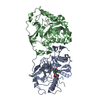
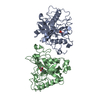
| Title | STRUCTURE OF INORGANIC PYROPHOSPHATASE | ||||||
 Components Components | INORGANIC PYROPHOSPHATASE | ||||||
 Keywords Keywords | HYDROLASE / PYROPHOSPHATE PHOSPHOHYDROLASE / MANGANESE | ||||||
| Function / homology |  Function and homology information Function and homology informationCytosolic tRNA aminoacylation / Mitochondrial tRNA aminoacylation / Pyrophosphate hydrolysis / inorganic diphosphatase / inorganic diphosphate phosphatase activity / phosphate-containing compound metabolic process / magnesium ion binding / nucleus / cytoplasm Similarity search - Function | ||||||
| Biological species |  | ||||||
| Method |  X-RAY DIFFRACTION / SYNCHROTRON / MOLECULAR REPLACEMENT / Resolution: 1.15 Å X-RAY DIFFRACTION / SYNCHROTRON / MOLECULAR REPLACEMENT / Resolution: 1.15 Å | ||||||
 Authors Authors | Heikinheimo, P. / Tuominen, V. / Ahonen, A.-K. / Teplyakov, A. / Cooperman, B.S. / Baykov, A.A. / Lahti, R. / Goldman, A. | ||||||
 Citation Citation | Journal: Proc.Natl.Acad.Sci.USA / Year: 2001 Title: Toward a Quantum-Mechanical Description of Metal-Assisted Phosphoryl Transfer in Pyrophosphatase Authors: Heikinheimo, P. / Tuominen, V. / Ahonen, A.-K. / Teplyakov, A. / Cooperman, B.S. / Baykov, A.A. / Lahti, R. / Goldman, A. | ||||||
| History |
| ||||||
| Remark 650 | HELIX DETERMINATION METHOD: AUTHOR PROVIDED. | ||||||
| Remark 700 | SHEET DETERMINATION METHOD: AUTHOR PROVIDED. |
- Structure visualization
| Structure viewer | Molecule: MolmilJmol/JSmol |
|---|
- Downloads & links
-Download
| PDBx/mmCIF format | 1e9g.cif.gz | 446.5 KB | Display | PDBx/mmCIF format |
|---|---|---|---|---|
| PDB format | pdb1e9g.ent.gz | 360.6 KB | Display | PDB format |
| PDBx/mmJSON format | 1e9g.json.gz | Tree view | PDBx/mmJSON format | |
| Others |  Other downloads Other downloads |
-Validation report
| Arichive directory | https://data.pdbj.org/pub/pdb/validation_reports/e9/1e9gftp://data.pdbj.org/pub/pdb/validation_reports/e9/1e9g | HTTPS FTP |
|---|
-Related structure data
| Related structure data |  1e6aC 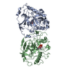 1wgjS C: citing same article ( S: Starting model for refinement |
|---|---|
| Similar structure data |


-Links
 PDBj
PDBj- Assembly
| Deposited unit | 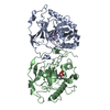
| ||||||||
|---|---|---|---|---|---|---|---|---|---|
| 1 |
| ||||||||
| Unit cell |
| ||||||||
| Noncrystallographic symmetry (NCS) | NCS oper: (Code: given Matrix: (0.7976, 0.6031, 0.015), Vector: |
-Components
| #1: Protein | Mass: 32225.375 Da / Num. of mol.: 2 Source method: isolated from a genetically manipulated source Details: PRODUCT COMPLEX Source: (gene. exp.) Cellular location: CYTOPLASM / Gene: PPA1 / Plasmid: PKW9 / Gene (production host): PPA1 / Production host:  #2: Chemical | ChemComp-MN /   Mass: 54.938 Da / Num. of mol.: 8 / Source method: obtained synthetically / Formula: Mn Mass: 54.938 Da / Num. of mol.: 8 / Source method: obtained synthetically / Formula: Mn#3: Chemical | ChemComp-PO4 /   Mass: 94.971 Da / Num. of mol.: 4 / Source method: obtained synthetically / Formula: PO4 Mass: 94.971 Da / Num. of mol.: 4 / Source method: obtained synthetically / Formula: PO4#4: Water | ChemComp-HOH / |  Mass: 18.015 Da / Num. of mol.: 1023 / Source method: isolated from a natural source / Formula: H2O Mass: 18.015 Da / Num. of mol.: 1023 / Source method: isolated from a natural source / Formula: H2OCompound details | IN PRESENCE OF DIVALENT METAL CATION CATALYSES THE REACTION: PYROPHOSPHATE + H(2)O = 2 ...IN PRESENCE OF DIVALENT METAL CATION CATALYSES THE REACTION: PYROPHOSPH | Sequence details | 1WGJ A SWS P00817 283 - 286 NOT IN ATOMS LIST 1WGJ B SWS P00817 283 - 286 NOT IN ATOMS LIST | |
|---|
-Experimental details
-Experiment
| Experiment | Method: X-RAY DIFFRACTION / Number of used crystals: 2 |
|---|
- Sample preparation
| Crystal | Density Matthews: 2.34 Å3/Da / Density % sol: 54.73 % Description: TWO MERGED DATA SETS: THE FIRST COLLECTED SEP-1996, WAVLENGTH 0.891 A, RESOLUTION RANGE 8-1.5 A AND THE SECOND COLLECTED 1997, WAVELENGTH 0.901 A, RESOLUTION RANGE 2.5-1.15 A | ||||||||||||||||||||||||||||||||||||
|---|---|---|---|---|---|---|---|---|---|---|---|---|---|---|---|---|---|---|---|---|---|---|---|---|---|---|---|---|---|---|---|---|---|---|---|---|---|
| Crystal grow | pH: 6 / Details: pH 6.00 | ||||||||||||||||||||||||||||||||||||
| Crystal grow | *PLUS Method: vapor diffusion, sitting drop / Details: Heikinheimo, P., (1996) Structure, 4, 1491. | ||||||||||||||||||||||||||||||||||||
| Components of the solutions | *PLUS
|
-Data collection
| Diffraction | Mean temperature: 100 K |
|---|---|
| Diffraction source | Source: SYNCHROTRON / Site: EMBL/DESY, HAMBURG  / Beamline: BW7A / Wavelength: 0.891 / Beamline: BW7A / Wavelength: 0.891 |
| Detector | Type: MARRESEARCH / Detector: IMAGE PLATE / Date: Sep 15, 1997 |
| Radiation | Protocol: SINGLE WAVELENGTH / Monochromatic (M) / Laue (L): M / Scattering type: x-ray |
| Radiation wavelength | Wavelength: 0.891 Å / Relative weight: 1 |
| Reflection | Resolution: 1.15→8 Å / Num. obs: 212495 / % possible obs: 85.4 % / Redundancy: 3.6 % / Rmerge(I) obs: 0.075 / Net I/σ(I): 11.95 |
| Reflection shell | Resolution: 1.15→1.17 Å / Rmerge(I) obs: 0.37 / Mean I/σ(I) obs: 3 / % possible all: 75.5 |
| Reflection shell | *PLUS % possible obs: 75.5 % |
- Processing
| Software |
| |||||||||||||||||||||||||||||||||
|---|---|---|---|---|---|---|---|---|---|---|---|---|---|---|---|---|---|---|---|---|---|---|---|---|---|---|---|---|---|---|---|---|---|---|
| Refinement | Method to determine structure: MOLECULAR REPLACEMENT Starting model: 1WGJ Resolution: 1.15→8 Å / Num. parameters: 51975 / Num. restraintsaints: 60843 / Cross valid method: FREE R-VALUE / σ(F): 0 / Stereochemistry target values: ENGH AND HUBER Details: ANISOTROPIC REFINEMENT REDUCED FREE R (NO CUTOFF) BY 4.7% THE C-TERMINAL RESIDUE WAS NOT SEEN IN THE DENSITY MAPS
| |||||||||||||||||||||||||||||||||
| Solvent computation | Solvent model: MOEWS & KRETSINGER, J.MOL.BIOL.91(1973)201-2 | |||||||||||||||||||||||||||||||||
| Refine analyze | Num. disordered residues: 42 / Occupancy sum hydrogen: 4448 / Occupancy sum non hydrogen: 5562.12 | |||||||||||||||||||||||||||||||||
| Refinement step | Cycle: LAST / Resolution: 1.15→8 Å
| |||||||||||||||||||||||||||||||||
| Refine LS restraints |
| |||||||||||||||||||||||||||||||||
| Software | *PLUS Name: CNS / Version: 0.5 / Classification: refinement | |||||||||||||||||||||||||||||||||
| Refine LS restraints | *PLUS
|
