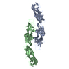
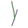
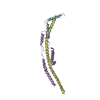
NetrinreceptorDCC / Colorectal cancer suppressor / Immunoglobulin superfamily DCC subclass member 1 / Tumor suppressor protein DCC
Mass: 12021.081 Da / Num. of mol.: 2 / Fragment: UNP residues 721-815 Source method: isolated from a genetically manipulated source Source: (gene. exp.) Homo sapiens (human) / Gene: DCC, IGDCC1 / Production host: Escherichia coli (E. coli) / References: UniProt: P43146
#2: Protein
NetrinreceptorDCC / Colorectal cancer suppressor / Immunoglobulin superfamily DCC subclass member 1 / Tumor suppressor protein DCC
Mass: 23357.670 Da / Num. of mol.: 2 / Fragment: UNP residues 844-1043 Source method: isolated from a genetically manipulated source Source: (gene. exp.) Homo sapiens (human) / Gene: DCC, IGDCC1 / Production host: Escherichia coli (E. coli) / References: UniProt: P43146
Compound details
Entities 1 and 2 are genetically manipulated as one entire chain. Hence, the structure contains 2 ...Entities 1 and 2 are genetically manipulated as one entire chain. Hence, the structure contains 2 molecules. However, residues 816-843 are disordered in both molecules. Currently each molecule is split into 2 chains in this structure. N-terminal domains are assigned chain IDs A/C, and C-terminal domains are assigned chain IDs B/D. Author are not certain whether chains A/B or A/D (C/D or C/B) belongs to one molecule.
-
Experimental details
-
Experiment
Experiment
Method: X-RAY DIFFRACTION / Number of used crystals: 1
-
Sample preparation
Crystal
Density Matthews: 2.7 Å3/Da / Density % sol: 54.49 %
In the structure databanks used in Yorodumi, some data are registered as the other names, "COVID-19 virus" and "2019-nCoV". Here are the details of the virus and the list of structure data.
Jan 31, 2019. EMDB accession codes are about to change! (news from PDBe EMDB page)
EMDB accession codes are about to change! (news from PDBe EMDB page)
The allocation of 4 digits for EMDB accession codes will soon come to an end. Whilst these codes will remain in use, new EMDB accession codes will include an additional digit and will expand incrementally as the available range of codes is exhausted. The current 4-digit format prefixed with “EMD-” (i.e. EMD-XXXX) will advance to a 5-digit format (i.e. EMD-XXXXX), and so on. It is currently estimated that the 4-digit codes will be depleted around Spring 2019, at which point the 5-digit format will come into force.
The EM Navigator/Yorodumi systems omit the EMD- prefix.
Related info.:Q: What is EMD? / ID/Accession-code notation in Yorodumi/EM Navigator
Yorodumi is a browser for structure data from EMDB, PDB, SASBDB, etc.
This page is also the successor to EM Navigator detail page, and also detail information page/front-end page for Omokage search.
The word "yorodu" (or yorozu) is an old Japanese word meaning "ten thousand". "mi" (miru) is to see.
Related info.:EMDB / PDB / SASBDB / Comparison of 3 databanks / Yorodumi Search / Aug 31, 2016. New EM Navigator & Yorodumi / Yorodumi Papers / Jmol/JSmol / Function and homology information / Changes in new EM Navigator and Yorodumi
 Movie
Movie Controller


 Open data
 Basic information
 Components
 Keywords
 Function and homology information
 Homo sapiens (human)
 X-RAY DIFFRACTION /
  Authors
 China, 2items
  Citation
 Structure visualization
 Downloads & links
 Other downloads
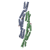


 PDBj
PDBj


 Assembly

 Sample preparation
 / Beamline: BL-1A / Wavelength: 1.1 Å
 Processing