 Movie
Movie Controller

[English] 日本語
 Yorodumi
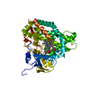
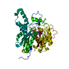
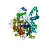
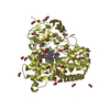
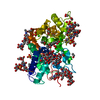
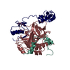
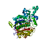
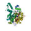
- PDB-5x7e: Crystal structure of vitamin D hydroxylase cytochrome P450 105A1 ... -
+ Open data

- Basic information
| Entry | Database: PDB / ID: 5x7e | ||||||
|---|---|---|---|---|---|---|---|
| Title | Crystal structure of vitamin D hydroxylase cytochrome P450 105A1 (R84A mutant) in complex with 1,25-dihydroxyvitamin D2 | ||||||
 Components Components | Vitamin D3 dihydroxylase | ||||||
 Keywords Keywords | OXIDOREDUCTASE / METAL-BINDING / MONOOXYGENASE | ||||||
| Function / homology |  Function and homology information Function and homology informationvitamin D 1,25-hydroxylase / vitamin D3 metabolic process / oxidoreductase activity, acting on paired donors, with incorporation or reduction of molecular oxygen, NAD(P)H as one donor, and incorporation of one atom of oxygen / iron ion binding / heme binding / cytoplasm Similarity search - Function | ||||||
| Biological species |  Streptomyces griseolus (bacteria) Streptomyces griseolus (bacteria) | ||||||
| Method |  X-RAY DIFFRACTION / SYNCHROTRON / MOLECULAR REPLACEMENT / Resolution: 1.9 Å X-RAY DIFFRACTION / SYNCHROTRON / MOLECULAR REPLACEMENT / Resolution: 1.9 Å | ||||||
| Model details | CYP105A1 in complex with active form of vitamin D2 | ||||||
 Authors Authors | Hayashi, K. / Yasuda, K. / Shiro, Y. / Sugimoto, H. / Sakaki, T. | ||||||
 Citation Citation | Journal: Biochem. Biophys. Res. Commun. / Year: 2017 Title: Production of an active form of vitamin D2 by genetically engineered CYP105A1 Authors: Yasuda, K. / Yogo, Y. / Sugimoto, H. / Mano, H. / Takita, T. / Ohta, M. / Kamakura, M. / Ikushiro, S. / Yasukawa, K. / Shiro, Y. / Sakaki, T. | ||||||
| History |
|
- Structure visualization
| Structure viewer | Molecule: MolmilJmol/JSmol |
|---|
- Downloads & links
-Download
| PDBx/mmCIF format | 5x7e.cif.gz | 100.9 KB | Display | PDBx/mmCIF format |
|---|---|---|---|---|
| PDB format | pdb5x7e.ent.gz | 74.7 KB | Display | PDB format |
| PDBx/mmJSON format | 5x7e.json.gz | Tree view | PDBx/mmJSON format | |
| Others |  Other downloads Other downloads |
-Validation report
| Arichive directory | https://data.pdbj.org/pub/pdb/validation_reports/x7/5x7eftp://data.pdbj.org/pub/pdb/validation_reports/x7/5x7e | HTTPS FTP |
|---|
-Related structure data
| Related structure data |  2zbzS S: Starting model for refinement |
|---|---|
| Similar structure data |

-Links
 PDBj
PDBj
- Assembly
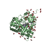
| Deposited unit | 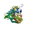
| ||||||||
|---|---|---|---|---|---|---|---|---|---|
| 1 |
| ||||||||
| Unit cell |
|
-Components
| #1: Protein | Mass: 44991.742 Da / Num. of mol.: 1 / Mutation: R84A Source method: isolated from a genetically manipulated source Source: (gene. exp.) Streptomyces griseolus (bacteria) / Gene: cyp105A1, suaC / Plasmid: pKK223-3 / Production host: References: UniProt: P18326, Oxidoreductases; Acting on paired donors, with incorporation or reduction of molecular oxygen; With reduced iron-sulfur protein as one donor, and incorporation of one ...References: UniProt: P18326, Oxidoreductases; Acting on paired donors, with incorporation or reduction of molecular oxygen; With reduced iron-sulfur protein as one donor, and incorporation of one atom of oxygen into the other donor |
|---|---|
| #2: Chemical | ChemComp-HEM /   Mass: 616.487 Da / Num. of mol.: 1 / Source method: obtained synthetically / Formula: C34H32FeN4O4 Mass: 616.487 Da / Num. of mol.: 1 / Source method: obtained synthetically / Formula: C34H32FeN4O4 |
| #3: Chemical | ChemComp-7ZU / (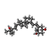  Mass: 428.647 Da / Num. of mol.: 1 / Source method: obtained synthetically / Formula: C28H44O3 Mass: 428.647 Da / Num. of mol.: 1 / Source method: obtained synthetically / Formula: C28H44O3 |
| #4: Water | ChemComp-HOH /  Mass: 18.015 Da / Num. of mol.: 294 / Source method: isolated from a natural source / Formula: H2O Mass: 18.015 Da / Num. of mol.: 294 / Source method: isolated from a natural source / Formula: H2O |
-Experimental details
-Experiment
| Experiment | Method: X-RAY DIFFRACTION / Number of used crystals: 1 |
|---|
- Sample preparation
| Crystal | Density Matthews: 2.17 Å3/Da / Density % sol: 43.27 % / Mosaicity: 0.59 ° |
|---|---|
| Crystal grow | Temperature: 283 K / Method: vapor diffusion / pH: 6.8 / Details: 26% PEGMME 20000, 0.2 M NaCl, 0.1 M BisTris |
-Data collection
| Diffraction | Mean temperature: 100 K |
|---|---|
| Diffraction source | Source: SYNCHROTRON / Site: SPring-8  / Beamline: BL26B2 / Wavelength: 0.97 Å / Beamline: BL26B2 / Wavelength: 0.97 Å |
| Detector | Type: RIGAKU JUPITER 210 / Detector: CCD / Date: Oct 10, 2008 / Details: mirrors |
| Radiation | Monochromator: Si(111) / Protocol: SINGLE WAVELENGTH / Monochromatic (M) / Laue (L): M / Scattering type: x-ray |
| Radiation wavelength | Wavelength: 0.97 Å / Relative weight: 1 |
| Reflection | Resolution: 1.9→20 Å / Num. obs: 30958 / % possible obs: 98 % / Redundancy: 7.2 % / Rmerge(I) obs: 0.062 / Net I/σ(I): 12 |
| Reflection shell | Resolution: 1.9→1.97 Å / Redundancy: 6.1 % / Rmerge(I) obs: 0.3 / % possible all: 89.2 |
- Processing
| Software |
| ||||||||||||||||||||||||||||||||||||||||||||||||||||||||||||||||||||||||||||||||||||||||||||||||||||||||||||||||||||||||||||||||||||||||||||||||||||||||||||||||||||||||||||||||||||||
|---|---|---|---|---|---|---|---|---|---|---|---|---|---|---|---|---|---|---|---|---|---|---|---|---|---|---|---|---|---|---|---|---|---|---|---|---|---|---|---|---|---|---|---|---|---|---|---|---|---|---|---|---|---|---|---|---|---|---|---|---|---|---|---|---|---|---|---|---|---|---|---|---|---|---|---|---|---|---|---|---|---|---|---|---|---|---|---|---|---|---|---|---|---|---|---|---|---|---|---|---|---|---|---|---|---|---|---|---|---|---|---|---|---|---|---|---|---|---|---|---|---|---|---|---|---|---|---|---|---|---|---|---|---|---|---|---|---|---|---|---|---|---|---|---|---|---|---|---|---|---|---|---|---|---|---|---|---|---|---|---|---|---|---|---|---|---|---|---|---|---|---|---|---|---|---|---|---|---|---|---|---|---|---|
| Refinement | Method to determine structure: MOLECULAR REPLACEMENT Starting model: 2ZBZ Resolution: 1.9→17.53 Å / Cor.coef. Fo:Fc: 0.95 / Cor.coef. Fo:Fc free: 0.915 / SU B: 3.426 / SU ML: 0.103 / Cross valid method: THROUGHOUT / σ(F): 0 / ESU R: 0.172 / ESU R Free: 0.155 / Details: HYDROGENS HAVE BEEN USED IF PRESENT IN
| ||||||||||||||||||||||||||||||||||||||||||||||||||||||||||||||||||||||||||||||||||||||||||||||||||||||||||||||||||||||||||||||||||||||||||||||||||||||||||||||||||||||||||||||||||||||
| Solvent computation | Ion probe radii: 0.8 Å / Shrinkage radii: 0.8 Å / VDW probe radii: 1.2 Å | ||||||||||||||||||||||||||||||||||||||||||||||||||||||||||||||||||||||||||||||||||||||||||||||||||||||||||||||||||||||||||||||||||||||||||||||||||||||||||||||||||||||||||||||||||||||
| Displacement parameters | Biso mean: 22.41 Å2
| ||||||||||||||||||||||||||||||||||||||||||||||||||||||||||||||||||||||||||||||||||||||||||||||||||||||||||||||||||||||||||||||||||||||||||||||||||||||||||||||||||||||||||||||||||||||
| Refinement step | Cycle: LAST / Resolution: 1.9→17.53 Å
| ||||||||||||||||||||||||||||||||||||||||||||||||||||||||||||||||||||||||||||||||||||||||||||||||||||||||||||||||||||||||||||||||||||||||||||||||||||||||||||||||||||||||||||||||||||||
| Refine LS restraints |
|