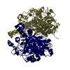
 Movie
Movie Controller


+ Open data

- Basic information
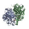
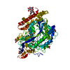
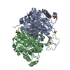
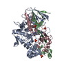
| Entry | Database: PDB / ID: 4c6u | ||||||
|---|---|---|---|---|---|---|---|
| Title | Crystal structure of M. tuberculosis KasA in complex with TLM5 | ||||||
 Components Components | 3-OXOACYL-[ACYL-CARRIER-PROTEIN] SYNTHASE 1 | ||||||
 Keywords Keywords | TRANSFERASE / KAS ENZYME / TYPE 2 FATTY ACID BIOSYNTHESIS / MYCOLIC ACID SYNTHESIS / THIOLACTOMYCIN | ||||||
| Function / homology | Thiolase/Chalcone synthase / Peroxisomal Thiolase; Chain A, domain 1 / 3-Layer(aba) Sandwich / Alpha Beta / : / Chem-TLG / :  Function and homology information Function and homology information | ||||||
| Biological species |   MYCOBACTERIUM TUBERCULOSIS (bacteria) MYCOBACTERIUM TUBERCULOSIS (bacteria) | ||||||
| Method |  X-RAY DIFFRACTION / SYNCHROTRON / MOLECULAR REPLACEMENT / Resolution: 2.4 Å X-RAY DIFFRACTION / SYNCHROTRON / MOLECULAR REPLACEMENT / Resolution: 2.4 Å | ||||||
 Authors Authors | Schiebel, J. / Kapilashrami, K. / Fekete, A. / Bommineni, G.R. / Schaefer, C.M. / Mueller, M.J. / Tonge, P.J. / Kisker, C. | ||||||
 Citation Citation | Journal: J.Biol.Chem. / Year: 2013 Title: Structural Basis for the Recognition of Mycolic Acid Precursors by Kasa, a Condensing Enzyme and Drug Target from Mycobacterium Tuberculosis Authors: Schiebel, J. / Kapilashrami, K. / Fekete, A. / Bommineni, G.R. / Schaefer, C.M. / Mueller, M.J. / Tonge, P.J. / Kisker, C. | ||||||
| History |
|
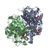
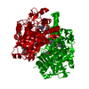
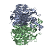
- Structure visualization
| Structure viewer | Molecule: MolmilJmol/JSmol |
|---|
- Downloads & links
-Download
| PDBx/mmCIF format | 4c6u.cif.gz | 171.1 KB | Display | PDBx/mmCIF format |
|---|---|---|---|---|
| PDB format | pdb4c6u.ent.gz | 135 KB | Display | PDB format |
| PDBx/mmJSON format | 4c6u.json.gz | Tree view | PDBx/mmJSON format | |
| Others |  Other downloads Other downloads |
-Validation report
| Arichive directory | https://data.pdbj.org/pub/pdb/validation_reports/c6/4c6uftp://data.pdbj.org/pub/pdb/validation_reports/c6/4c6u | HTTPS FTP |
|---|
-Related structure data
| Related structure data | 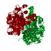 4c6vC 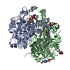 4c6wC 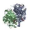 4c6xC 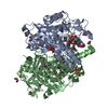 4c6zC  4c70C 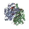 4c71C 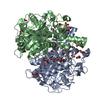 4c72C 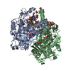 4c73C 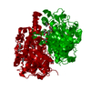 2wgeS C: citing same article ( S: Starting model for refinement |
|---|---|
| Similar structure data |


-Links
 PDBj
PDBj- Assembly
| Deposited unit | 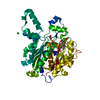
| ||||||||
|---|---|---|---|---|---|---|---|---|---|
| 1 | 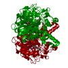
| ||||||||
| Unit cell |
| ||||||||
| Components on special symmetry positions |
|
-Components
-Protein , 1 types, 1 molecules A
| #1: Protein | Mass: 45820.688 Da / Num. of mol.: 1 Source method: isolated from a genetically manipulated source Source: (gene. exp.) MYCOBACTERIUM TUBERCULOSIS (bacteria) / Strain: H37RV / Plasmid: PFPCA1 / Production host: MYCOBACTERIUM SMEGMATIS (bacteria) / Strain (production host): MC2155References: UniProt: I6Y8T4, beta-ketoacyl-[acyl-carrier-protein] synthase I |
|---|
-Non-polymers , 5 types, 119 molecules 


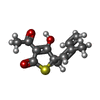

| #2: Chemical |  Mass: 62.068 Da / Num. of mol.: 3 / Source method: obtained synthetically / Formula: C2H6O2 Mass: 62.068 Da / Num. of mol.: 3 / Source method: obtained synthetically / Formula: C2H6O2#3: Chemical | ChemComp-CL / |  Mass: 35.453 Da / Num. of mol.: 1 / Source method: obtained synthetically / Formula: Cl Mass: 35.453 Da / Num. of mol.: 1 / Source method: obtained synthetically / Formula: Cl#4: Chemical | ChemComp-K / |  Mass: 39.098 Da / Num. of mol.: 1 / Source method: obtained synthetically / Formula: K Mass: 39.098 Da / Num. of mol.: 1 / Source method: obtained synthetically / Formula: K#5: Chemical | ChemComp-TLG / ( |  Mass: 238.303 Da / Num. of mol.: 1 / Source method: obtained synthetically / Formula: C12H14O3S Mass: 238.303 Da / Num. of mol.: 1 / Source method: obtained synthetically / Formula: C12H14O3S#6: Water | ChemComp-HOH / | Mass: 18.015 Da / Num. of mol.: 113 / Source method: isolated from a natural source / Formula: H2O |
|---|
-Experimental details
-Experiment
| Experiment | Method: X-RAY DIFFRACTION / Number of used crystals: 1 |
|---|
- Sample preparation
| Crystal | Density Matthews: 2.96 Å3/Da / Density % sol: 58.47 % / Description: NONE |
|---|---|
| Crystal grow | pH: 9.5 Details: 10% PEG 3350, 0.2 M K/NA-TARTRATE, 1.5 MM TCEP, pH 9.5 |
-Data collection
| Diffraction | Mean temperature: 100 K |
|---|---|
| Diffraction source | Source: SYNCHROTRON / Site: ESRF  / Beamline: ID23-2 / Wavelength: 0.8726 / Beamline: ID23-2 / Wavelength: 0.8726 |
| Detector | Type: ADSC CCD / Detector: CCD / Date: Oct 11, 2012 |
| Radiation | Protocol: SINGLE WAVELENGTH / Monochromatic (M) / Laue (L): M / Scattering type: x-ray |
| Radiation wavelength | Wavelength: 0.8726 Å / Relative weight: 1 |
| Reflection | Resolution: 2.4→49.22 Å / Num. obs: 20763 / % possible obs: 99.8 % / Observed criterion σ(I): 6 / Redundancy: 7.1 % / Rmerge(I) obs: 0.1 / Net I/σ(I): 13.9 |
| Reflection shell | Resolution: 2.4→2.53 Å / Redundancy: 6 % / Rmerge(I) obs: 0.6 / Mean I/σ(I) obs: 2.7 / % possible all: 99.9 |
- Processing
| Software |
| ||||||||||||||||||||||||||||||||||||||||||||||||||||||||||||||||||||||||||||||||||||||||||||||||||||||||||||||||||||||||||||||||||||||||||||||||||||||||||||||||||||||||||||||||||||||
|---|---|---|---|---|---|---|---|---|---|---|---|---|---|---|---|---|---|---|---|---|---|---|---|---|---|---|---|---|---|---|---|---|---|---|---|---|---|---|---|---|---|---|---|---|---|---|---|---|---|---|---|---|---|---|---|---|---|---|---|---|---|---|---|---|---|---|---|---|---|---|---|---|---|---|---|---|---|---|---|---|---|---|---|---|---|---|---|---|---|---|---|---|---|---|---|---|---|---|---|---|---|---|---|---|---|---|---|---|---|---|---|---|---|---|---|---|---|---|---|---|---|---|---|---|---|---|---|---|---|---|---|---|---|---|---|---|---|---|---|---|---|---|---|---|---|---|---|---|---|---|---|---|---|---|---|---|---|---|---|---|---|---|---|---|---|---|---|---|---|---|---|---|---|---|---|---|---|---|---|---|---|---|---|
| Refinement | Method to determine structure: MOLECULAR REPLACEMENT Starting model: PDB ENTRY 2WGE Resolution: 2.4→67.19 Å / Cor.coef. Fo:Fc: 0.961 / Cor.coef. Fo:Fc free: 0.937 / SU B: 14.493 / SU ML: 0.153 / Cross valid method: THROUGHOUT / ESU R: 0.27 / ESU R Free: 0.213 / Stereochemistry target values: MAXIMUM LIKELIHOOD / Details: HYDROGENS HAVE BEEN ADDED IN THE RIDING POSITIONS.
| ||||||||||||||||||||||||||||||||||||||||||||||||||||||||||||||||||||||||||||||||||||||||||||||||||||||||||||||||||||||||||||||||||||||||||||||||||||||||||||||||||||||||||||||||||||||
| Solvent computation | Ion probe radii: 0.8 Å / Shrinkage radii: 0.8 Å / VDW probe radii: 1.4 Å / Solvent model: BABINET MODEL WITH MASK | ||||||||||||||||||||||||||||||||||||||||||||||||||||||||||||||||||||||||||||||||||||||||||||||||||||||||||||||||||||||||||||||||||||||||||||||||||||||||||||||||||||||||||||||||||||||
| Displacement parameters | Biso mean: 52.186 Å2
| ||||||||||||||||||||||||||||||||||||||||||||||||||||||||||||||||||||||||||||||||||||||||||||||||||||||||||||||||||||||||||||||||||||||||||||||||||||||||||||||||||||||||||||||||||||||
| Refinement step | Cycle: LAST / Resolution: 2.4→67.19 Å
| ||||||||||||||||||||||||||||||||||||||||||||||||||||||||||||||||||||||||||||||||||||||||||||||||||||||||||||||||||||||||||||||||||||||||||||||||||||||||||||||||||||||||||||||||||||||
| Refine LS restraints |
|