 Movie
Movie Controller

[English] 日本語
 Yorodumi
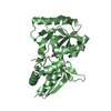
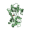
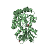
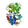
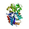
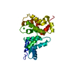
- PDB-5x3h: The Y81G mutant of the UNG crystal structure from Nitratifractor ... -
+ Open data

- Basic information
| Entry | Database: PDB / ID: 5x3h | |||||||||
|---|---|---|---|---|---|---|---|---|---|---|
| Title | The Y81G mutant of the UNG crystal structure from Nitratifractor salsuginis | |||||||||
 Components Components | Uracil-DNA glycosylase | |||||||||
 Keywords Keywords | DNA BINDING PROTEIN / uracil DNA glycosylase / base excision repair | |||||||||
| Function / homology | base-excision repair, AP site formation via deaminated base removal / Uracil-DNA glycosylase family 1 / Uracil-DNA glycosylase-like domain superfamily / uracil DNA N-glycosylase activity / Uracil-DNA glycosylase Function and homology information Function and homology information | |||||||||
| Biological species |  Nitratifractor salsuginis (bacteria) Nitratifractor salsuginis (bacteria) | |||||||||
| Method |  X-RAY DIFFRACTION / SYNCHROTRON / MOLECULAR REPLACEMENT / Resolution: 2.5 Å X-RAY DIFFRACTION / SYNCHROTRON / MOLECULAR REPLACEMENT / Resolution: 2.5 Å | |||||||||
 Authors Authors | Xie, W. / Chen, R. / Cao, W. / Zhang, Z. | |||||||||
| Funding support |  China, 2items China, 2items
| |||||||||
 Citation Citation | Journal: FEBS J. / Year: 2017 Title: An unconventional family 1 uracil DNA glycosylase in Nitratifractor salsuginis. Authors: Li, J. / Chen, R. / Yang, Y. / Zhang, Z. / Fang, G.C. / Xie, W. / Cao, W. | |||||||||
| History |
|
- Structure visualization
| Structure viewer | Molecule: MolmilJmol/JSmol |
|---|
- Downloads & links
-Download
| PDBx/mmCIF format | 5x3h.cif.gz | 107.4 KB | Display | PDBx/mmCIF format |
|---|---|---|---|---|
| PDB format | pdb5x3h.ent.gz | 81 KB | Display | PDB format |
| PDBx/mmJSON format | 5x3h.json.gz | Tree view | PDBx/mmJSON format | |
| Others |  Other downloads Other downloads |
-Validation report
| Summary document | 5x3h_validation.pdf.gz | 432.1 KB | Display | wwPDB validaton report |
|---|---|---|---|---|
| Full document | 5x3h_full_validation.pdf.gz | 435.9 KB | Display | |
| Data in XML | 5x3h_validation.xml.gz | 18.5 KB | Display | |
| Data in CIF | 5x3h_validation.cif.gz | 25.1 KB | Display | |
| Arichive directory | https://data.pdbj.org/pub/pdb/validation_reports/x3/5x3hftp://data.pdbj.org/pub/pdb/validation_reports/x3/5x3h | HTTPS FTP |
-Related structure data
| Related structure data | 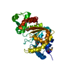 5x3gSC S: Starting model for refinement C: citing same article ( |
|---|---|
| Similar structure data |


-Links
 PDBj
PDBj- Assembly
| Deposited unit | 
| ||||||||
|---|---|---|---|---|---|---|---|---|---|
| 1 | 
| ||||||||
| 2 | 
| ||||||||
| Unit cell |
|
-Components
| #1: Protein | Mass: 30398.912 Da / Num. of mol.: 2 / Mutation: Y81G Source method: isolated from a genetically manipulated source Source: (gene. exp.) Nitratifractor salsuginis (strain DSM 16511 / JCM 12458 / E9I37-1) (bacteria)Strain: DSM 16511 / JCM 12458 / E9I37-1 / Gene: Nitsa_0175 / Production host: #2: Water | ChemComp-HOH / |  Mass: 18.015 Da / Num. of mol.: 24 / Source method: isolated from a natural source / Formula: H2O Mass: 18.015 Da / Num. of mol.: 24 / Source method: isolated from a natural source / Formula: H2OHas protein modification | Y | |
|---|
-Experimental details
-Experiment
| Experiment | Method: X-RAY DIFFRACTION / Number of used crystals: 1 |
|---|
- Sample preparation
| Crystal | Density Matthews: 2.01 Å3/Da / Density % sol: 38.89 % |
|---|---|
| Crystal grow | Temperature: 298 K / Method: vapor diffusion, sitting drop / pH: 7 / Details: 10% PEG3350 ?0.1 M HEPES pH7.0 |
-Data collection
| Diffraction | Mean temperature: 100 K |
|---|---|
| Diffraction source | Source: SYNCHROTRON / Site: SSRF / Beamline: BL17U / Wavelength: 0.99 Å |
| Detector | Type: ADSC QUANTUM 315r / Detector: CCD / Date: Apr 2, 2016 |
| Radiation | Protocol: SINGLE WAVELENGTH / Monochromatic (M) / Laue (L): M / Scattering type: x-ray |
| Radiation wavelength | Wavelength: 0.99 Å / Relative weight: 1 |
| Reflection | Resolution: 2.5→50 Å / Num. obs: 16628 / % possible obs: 99.2 % / Redundancy: 3.5 % / Rmerge(I) obs: 0.079 / Net I/σ(I): 16.5 |
| Reflection shell | Resolution: 2.5→2.59 Å / Redundancy: 3.4 % / Rmerge(I) obs: 0.263 / Mean I/σ(I) obs: 5.8 / Num. unique obs: 1623 / CC1/2: 0.92 / % possible all: 98.5 |
- Processing
| Software |
| ||||||||||||||||||||||||||||||||||||||||||
|---|---|---|---|---|---|---|---|---|---|---|---|---|---|---|---|---|---|---|---|---|---|---|---|---|---|---|---|---|---|---|---|---|---|---|---|---|---|---|---|---|---|---|---|
| Refinement | Method to determine structure: MOLECULAR REPLACEMENT Starting model: 5X3G Resolution: 2.5→39.9 Å / SU ML: 0.41 / Cross valid method: FREE R-VALUE / σ(F): 1.39 / Phase error: 29.41
| ||||||||||||||||||||||||||||||||||||||||||
| Solvent computation | Shrinkage radii: 0.9 Å / VDW probe radii: 1.11 Å | ||||||||||||||||||||||||||||||||||||||||||
| Refinement step | Cycle: LAST / Resolution: 2.5→39.9 Å
| ||||||||||||||||||||||||||||||||||||||||||
| Refine LS restraints |
| ||||||||||||||||||||||||||||||||||||||||||
| LS refinement shell |
|
