 Movie
Movie Controller

[English] 日本語
 Yorodumi
- PDB-2yjp: Crystal structure of the solute receptors for L-cysteine of Neiss... -
+ Open data

- Basic information
| Entry | Database: PDB / ID: 2yjp | ||||||
|---|---|---|---|---|---|---|---|
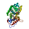
| Title | Crystal structure of the solute receptors for L-cysteine of Neisseria gonorrhoeae | ||||||
 Components Components | PUTATIVE ABC TRANSPORTER, PERIPLASMIC BINDING PROTEIN, AMINO ACID | ||||||
 Keywords Keywords | TRANSPORT PROTEIN / SOLUTE-BINDING PROTEIN | ||||||
| Function / homology |  Function and homology information Function and homology informationamino acid transport / outer membrane-bounded periplasmic space / extracellular region / metal ion binding Similarity search - Function | ||||||
| Biological species |  NEISSERIA GONORRHOEAE (bacteria) NEISSERIA GONORRHOEAE (bacteria) | ||||||
| Method |  X-RAY DIFFRACTION / SYNCHROTRON / MOLECULAR REPLACEMENT / Resolution: 2.26 Å X-RAY DIFFRACTION / SYNCHROTRON / MOLECULAR REPLACEMENT / Resolution: 2.26 Å | ||||||
 Authors Authors | Bulut, H. / Moniot, S. / Scheffel, F. / Gathmann, S. / Licht, A. / Saenger, W. / Schneider, E. | ||||||
 Citation Citation | Journal: J.Mol.Biol. / Year: 2012 Title: Crystal Structures of Two Solute Receptors for L-Cystine and L-Cysteine, Respectively, of the Human Pathogen Neisseria Gonorrhoeae. Authors: Bulut, H. / Moniot, S. / Licht, A. / Scheffel, F. / Gathmann, S. / Saenger, W. / Schneider, E. | ||||||
| History |
|
- Structure visualization
| Structure viewer | Molecule: MolmilJmol/JSmol |
|---|
- Downloads & links
-Download
| PDBx/mmCIF format | 2yjp.cif.gz | 302.1 KB | Display | PDBx/mmCIF format |
|---|---|---|---|---|
| PDB format | pdb2yjp.ent.gz | 244 KB | Display | PDB format |
| PDBx/mmJSON format | 2yjp.json.gz | Tree view | PDBx/mmJSON format | |
| Others |  Other downloads Other downloads |
-Validation report
| Arichive directory | https://data.pdbj.org/pub/pdb/validation_reports/yj/2yjpftp://data.pdbj.org/pub/pdb/validation_reports/yj/2yjp | HTTPS FTP |
|---|
-Related structure data
| Related structure data | 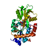 2ylnC  3zsfC  1xt8S C: citing same article ( S: Starting model for refinement |
|---|---|
| Similar structure data |


-Links
 PDBj
PDBj


- Assembly
| Deposited unit | 
| ||||||||||||||||||||||||||||||||||||||||||||||||||||||||||||||||||||||||||||||||||||||||||||||||||||||||||||||||||||||||||||||||||||||||||||||||||||||||||||||||||||||||||||||||||||||||||||||||||||||
|---|---|---|---|---|---|---|---|---|---|---|---|---|---|---|---|---|---|---|---|---|---|---|---|---|---|---|---|---|---|---|---|---|---|---|---|---|---|---|---|---|---|---|---|---|---|---|---|---|---|---|---|---|---|---|---|---|---|---|---|---|---|---|---|---|---|---|---|---|---|---|---|---|---|---|---|---|---|---|---|---|---|---|---|---|---|---|---|---|---|---|---|---|---|---|---|---|---|---|---|---|---|---|---|---|---|---|---|---|---|---|---|---|---|---|---|---|---|---|---|---|---|---|---|---|---|---|---|---|---|---|---|---|---|---|---|---|---|---|---|---|---|---|---|---|---|---|---|---|---|---|---|---|---|---|---|---|---|---|---|---|---|---|---|---|---|---|---|---|---|---|---|---|---|---|---|---|---|---|---|---|---|---|---|---|---|---|---|---|---|---|---|---|---|---|---|---|---|---|---|
| 1 | 
| ||||||||||||||||||||||||||||||||||||||||||||||||||||||||||||||||||||||||||||||||||||||||||||||||||||||||||||||||||||||||||||||||||||||||||||||||||||||||||||||||||||||||||||||||||||||||||||||||||||||
| 2 | 
| ||||||||||||||||||||||||||||||||||||||||||||||||||||||||||||||||||||||||||||||||||||||||||||||||||||||||||||||||||||||||||||||||||||||||||||||||||||||||||||||||||||||||||||||||||||||||||||||||||||||
| 3 | 
| ||||||||||||||||||||||||||||||||||||||||||||||||||||||||||||||||||||||||||||||||||||||||||||||||||||||||||||||||||||||||||||||||||||||||||||||||||||||||||||||||||||||||||||||||||||||||||||||||||||||
| Unit cell |
| ||||||||||||||||||||||||||||||||||||||||||||||||||||||||||||||||||||||||||||||||||||||||||||||||||||||||||||||||||||||||||||||||||||||||||||||||||||||||||||||||||||||||||||||||||||||||||||||||||||||
| Noncrystallographic symmetry (NCS) | NCS domain:
NCS domain segments:
|
-Components
| #1: Protein | Mass: 31403.377 Da / Num. of mol.: 3 / Fragment: RESIDUES 18-284 / Mutation: YES Source method: isolated from a genetically manipulated source Source: (gene. exp.) NEISSERIA GONORRHOEAE (bacteria) / Strain: FA 1090 / Production host: #2: Chemical |   Type: L-peptide linking / Mass: 121.158 Da / Num. of mol.: 3 / Source method: obtained synthetically / Formula: C3H7NO2S Type: L-peptide linking / Mass: 121.158 Da / Num. of mol.: 3 / Source method: obtained synthetically / Formula: C3H7NO2S#3: Chemical | ChemComp-ZN /   Mass: 65.409 Da / Num. of mol.: 8 / Source method: obtained synthetically / Formula: Zn Mass: 65.409 Da / Num. of mol.: 8 / Source method: obtained synthetically / Formula: Zn#4: Chemical | ChemComp-EDO /   Mass: 62.068 Da / Num. of mol.: 6 / Source method: obtained synthetically / Formula: C2H6O2 Mass: 62.068 Da / Num. of mol.: 6 / Source method: obtained synthetically / Formula: C2H6O2#5: Water | ChemComp-HOH / |  Mass: 18.015 Da / Num. of mol.: 356 / Source method: isolated from a natural source / Formula: H2O Mass: 18.015 Da / Num. of mol.: 356 / Source method: isolated from a natural source / Formula: H2OCompound details | ENGINEERED RESIDUE IN CHAIN A, CYS 23 TO ALA ENGINEERED RESIDUE IN CHAIN B, CYS 23 TO ALA ...ENGINEERED | Sequence details | N-TERMINAL HIS-TAG CONSTRUCT WITH ENTEROKINA | |
|---|
-Experimental details
-Experiment
| Experiment | Method: X-RAY DIFFRACTION / Number of used crystals: 1 |
|---|
- Sample preparation
| Crystal | Density Matthews: 2.5 Å3/Da / Density % sol: 50.5 % / Description: NONE |
|---|---|
| Crystal grow | Details: 16-18% PEG 4000, 100 MM HEPES PH 7.0, 10 MM ZNCL2. |
-Data collection
| Diffraction | Mean temperature: 100 K |
|---|---|
| Diffraction source | Source: SYNCHROTRON / Site: BESSY  / Beamline: 14.1 / Wavelength: 0.9395 / Beamline: 14.1 / Wavelength: 0.9395 |
| Detector | Type: MARMOSAIC 225 mm CCD / Detector: CCD / Date: Jul 20, 2009 |
| Radiation | Protocol: SINGLE WAVELENGTH / Monochromatic (M) / Laue (L): M / Scattering type: x-ray |
| Radiation wavelength | Wavelength: 0.9395 Å / Relative weight: 1 |
| Reflection | Resolution: 2.26→47.18 Å / Num. obs: 39913 / % possible obs: 97.8 % / Observed criterion σ(I): -3 / Redundancy: 4.14 % / Biso Wilson estimate: 30.5 Å2 / Rmerge(I) obs: 0.11 / Net I/σ(I): 11.32 |
| Reflection shell | Resolution: 2.26→2.38 Å / Redundancy: 2.92 % / Rmerge(I) obs: 0.49 / Mean I/σ(I) obs: 2.25 / % possible all: 86.7 |
- Processing
| Software |
| ||||||||||||||||||||||||||||||||||||||||||||||||||||||||||||||||||||||||||||||||||||||||||||||||||||||||||||||||||||||||||||||||||||||||||||||||||||||||||||||||||||||||||||||||||||||
|---|---|---|---|---|---|---|---|---|---|---|---|---|---|---|---|---|---|---|---|---|---|---|---|---|---|---|---|---|---|---|---|---|---|---|---|---|---|---|---|---|---|---|---|---|---|---|---|---|---|---|---|---|---|---|---|---|---|---|---|---|---|---|---|---|---|---|---|---|---|---|---|---|---|---|---|---|---|---|---|---|---|---|---|---|---|---|---|---|---|---|---|---|---|---|---|---|---|---|---|---|---|---|---|---|---|---|---|---|---|---|---|---|---|---|---|---|---|---|---|---|---|---|---|---|---|---|---|---|---|---|---|---|---|---|---|---|---|---|---|---|---|---|---|---|---|---|---|---|---|---|---|---|---|---|---|---|---|---|---|---|---|---|---|---|---|---|---|---|---|---|---|---|---|---|---|---|---|---|---|---|---|---|---|
| Refinement | Method to determine structure: MOLECULAR REPLACEMENT Starting model: PDB ENTRY 1XT8 Resolution: 2.26→47.18 Å / Cor.coef. Fo:Fc: 0.94 / Cor.coef. Fo:Fc free: 0.911 / Cross valid method: THROUGHOUT / ESU R: 0.284 / ESU R Free: 0.214 / Stereochemistry target values: MAXIMUM LIKELIHOOD / Details: HYDROGENS HAVE BEEN ADDED IN THE RIDING POSITIONS.
| ||||||||||||||||||||||||||||||||||||||||||||||||||||||||||||||||||||||||||||||||||||||||||||||||||||||||||||||||||||||||||||||||||||||||||||||||||||||||||||||||||||||||||||||||||||||
| Solvent computation | Ion probe radii: 0.8 Å / Shrinkage radii: 0.8 Å / VDW probe radii: 1.4 Å / Solvent model: MASK | ||||||||||||||||||||||||||||||||||||||||||||||||||||||||||||||||||||||||||||||||||||||||||||||||||||||||||||||||||||||||||||||||||||||||||||||||||||||||||||||||||||||||||||||||||||||
| Displacement parameters | Biso mean: 26.081 Å2
| ||||||||||||||||||||||||||||||||||||||||||||||||||||||||||||||||||||||||||||||||||||||||||||||||||||||||||||||||||||||||||||||||||||||||||||||||||||||||||||||||||||||||||||||||||||||
| Refinement step | Cycle: LAST / Resolution: 2.26→47.18 Å
| ||||||||||||||||||||||||||||||||||||||||||||||||||||||||||||||||||||||||||||||||||||||||||||||||||||||||||||||||||||||||||||||||||||||||||||||||||||||||||||||||||||||||||||||||||||||
| Refine LS restraints |
|