 Movie
Movie Controller


+ Open data

- Basic information
| Entry | Database: PDB / ID: 5wxi | |||||||||
|---|---|---|---|---|---|---|---|---|---|---|
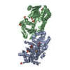
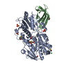
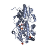
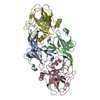
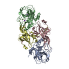
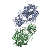
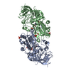
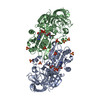
| Title | EarP bound with dTDP-rhamnose (soaked) | |||||||||
 Components Components | EarP | |||||||||
 Keywords Keywords | TRANSFERASE / glycosyltransferase / GT-B / EF-P / rhamnosylation / translation elongation / dTDP-rhamnose | |||||||||
| Function / homology | protein-arginine rhamnosyltransferase activity / Protein-arginine rhamnosyltransferase EarP / Elongation-Factor P (EF-P) rhamnosyltransferase EarP / Transferases; Glycosyltransferases; Hexosyltransferases / BETA-MERCAPTOETHANOL / 2'-DEOXY-THYMIDINE-BETA-L-RHAMNOSE / Protein-arginine rhamnosyltransferase Function and homology information Function and homology information | |||||||||
| Biological species | Neisseria meningitidis serogroup B / serotype 15 | |||||||||
| Method |  X-RAY DIFFRACTION / SYNCHROTRON / FOURIER SYNTHESIS / Resolution: 2 Å X-RAY DIFFRACTION / SYNCHROTRON / FOURIER SYNTHESIS / Resolution: 2 Å | |||||||||
 Authors Authors | Sengoku, T. / Yokoyama, S. / Yanagisawa, T. | |||||||||
| Funding support |  Japan, 2items Japan, 2items
| |||||||||
 Citation Citation | Journal: Nat. Chem. Biol. / Year: 2018 Title: Structural basis of protein arginine rhamnosylation by glycosyltransferase EarP Authors: Sengoku, T. / Suzuki, T. / Dohmae, N. / Watanabe, C. / Honma, T. / Hikida, Y. / Yamaguchi, Y. / Takahashi, H. / Yokoyama, S. / Yanagisawa, T. | |||||||||
| History |
|
- Structure visualization
| Structure viewer | Molecule: MolmilJmol/JSmol |
|---|
- Downloads & links
-Download
| PDBx/mmCIF format | 5wxi.cif.gz | 333.3 KB | Display | PDBx/mmCIF format |
|---|---|---|---|---|
| PDB format | pdb5wxi.ent.gz | 270.4 KB | Display | PDB format |
| PDBx/mmJSON format | 5wxi.json.gz | Tree view | PDBx/mmJSON format | |
| Others |  Other downloads Other downloads |
-Validation report
| Arichive directory | https://data.pdbj.org/pub/pdb/validation_reports/wx/5wxiftp://data.pdbj.org/pub/pdb/validation_reports/wx/5wxi | HTTPS FTP |
|---|
-Related structure data


-Links
 PDBj
PDBj


- Assembly
| Deposited unit | 
| ||||||||
|---|---|---|---|---|---|---|---|---|---|
| 1 |
| ||||||||
| Unit cell |
|
-Components
-Protein , 1 types, 2 molecules AB
| #1: Protein | Mass: 44219.992 Da / Num. of mol.: 2 Source method: isolated from a genetically manipulated source Source: (gene. exp.)  Neisseria meningitidis serogroup B / serotype 15 (strain H44/76) (bacteria) Neisseria meningitidis serogroup B / serotype 15 (strain H44/76) (bacteria)Strain: H44/76 / Gene: NMH_0797 / Plasmid: pET28c / Production host: |
|---|
-Non-polymers , 5 types, 651 molecules 


| #2: Chemical |  Mass: 548.330 Da / Num. of mol.: 2 / Source method: obtained synthetically / Formula: C16H26N2O15P2 Mass: 548.330 Da / Num. of mol.: 2 / Source method: obtained synthetically / Formula: C16H26N2O15P2#3: Chemical | ChemComp-BME /  Mass: 78.133 Da / Num. of mol.: 6 / Source method: obtained synthetically / Formula: C2H6OS Mass: 78.133 Da / Num. of mol.: 6 / Source method: obtained synthetically / Formula: C2H6OS#4: Chemical | ChemComp-SO4 /  Mass: 96.063 Da / Num. of mol.: 7 / Source method: obtained synthetically / Formula: SO4 Mass: 96.063 Da / Num. of mol.: 7 / Source method: obtained synthetically / Formula: SO4#5: Chemical | ChemComp-GOL / |  Mass: 92.094 Da / Num. of mol.: 1 / Source method: obtained synthetically / Formula: C3H8O3 Mass: 92.094 Da / Num. of mol.: 1 / Source method: obtained synthetically / Formula: C3H8O3#6: Water | ChemComp-HOH / | Mass: 18.015 Da / Num. of mol.: 635 / Source method: isolated from a natural source / Formula: H2O |
|---|
-Experimental details
-Experiment
| Experiment | Method: X-RAY DIFFRACTION / Number of used crystals: 1 |
|---|
- Sample preparation
| Crystal | Density Matthews: 2.5 Å3/Da / Density % sol: 50.78 % |
|---|---|
| Crystal grow | Temperature: 277 K / Method: vapor diffusion, sitting drop Details: 0.1M Hepes-NaOH buffer (pH 6.8-7.5), 20% PEG3350, 0.2M ammonium sulfate PH range: 6.8-7.5 |
-Data collection
| Diffraction | Mean temperature: 90 K |
|---|---|
| Diffraction source | Source: SYNCHROTRON / Site: Photon Factory / Beamline: BL-5A / Wavelength: 1 Å |
| Detector | Type: ADSC QUANTUM 315 / Detector: CCD / Date: Feb 23, 2016 |
| Radiation | Protocol: SINGLE WAVELENGTH / Monochromatic (M) / Laue (L): M / Scattering type: x-ray |
| Radiation wavelength | Wavelength: 1 Å / Relative weight: 1 |
| Reflection | Resolution: 2→48.85 Å / Num. obs: 61063 / % possible obs: 100 % / Redundancy: 7.3 % / Net I/σ(I): 10.7 |
| Reflection shell | Resolution: 2→2.05 Å / Redundancy: 7.2 % / Mean I/σ(I) obs: 1.9 / CC1/2: 0.649 / % possible all: 99.7 |
- Processing
| Software |
| |||||||||||||||||||||||||||||||||||||||||||||||||||||||||||||||||||||||||||||||||||||||||||||||||||||||||||||||||||||||||||||
|---|---|---|---|---|---|---|---|---|---|---|---|---|---|---|---|---|---|---|---|---|---|---|---|---|---|---|---|---|---|---|---|---|---|---|---|---|---|---|---|---|---|---|---|---|---|---|---|---|---|---|---|---|---|---|---|---|---|---|---|---|---|---|---|---|---|---|---|---|---|---|---|---|---|---|---|---|---|---|---|---|---|---|---|---|---|---|---|---|---|---|---|---|---|---|---|---|---|---|---|---|---|---|---|---|---|---|---|---|---|---|---|---|---|---|---|---|---|---|---|---|---|---|---|---|---|---|
| Refinement | Method to determine structure: FOURIER SYNTHESIS Starting model: apo EarP Resolution: 2→48.847 Å / SU ML: 0.19 / Cross valid method: FREE R-VALUE / σ(F): 1.35 / Phase error: 20.11
| |||||||||||||||||||||||||||||||||||||||||||||||||||||||||||||||||||||||||||||||||||||||||||||||||||||||||||||||||||||||||||||
| Solvent computation | Shrinkage radii: 0.9 Å / VDW probe radii: 1.11 Å | |||||||||||||||||||||||||||||||||||||||||||||||||||||||||||||||||||||||||||||||||||||||||||||||||||||||||||||||||||||||||||||
| Refinement step | Cycle: LAST / Resolution: 2→48.847 Å
| |||||||||||||||||||||||||||||||||||||||||||||||||||||||||||||||||||||||||||||||||||||||||||||||||||||||||||||||||||||||||||||
| Refine LS restraints |
| |||||||||||||||||||||||||||||||||||||||||||||||||||||||||||||||||||||||||||||||||||||||||||||||||||||||||||||||||||||||||||||
| LS refinement shell |
| |||||||||||||||||||||||||||||||||||||||||||||||||||||||||||||||||||||||||||||||||||||||||||||||||||||||||||||||||||||||||||||
| Refinement TLS params. | Method: refined / Refine-ID: X-RAY DIFFRACTION
| |||||||||||||||||||||||||||||||||||||||||||||||||||||||||||||||||||||||||||||||||||||||||||||||||||||||||||||||||||||||||||||
| Refinement TLS group |
|
