 Movie
Movie Controller

[English] 日本語
 Yorodumi
- PDB-5ws3: Crystal structures of human orexin 2 receptor bound to the select... -
+ Open data

- Basic information
| Entry | Database: PDB / ID: 5ws3 | |||||||||||||||
|---|---|---|---|---|---|---|---|---|---|---|---|---|---|---|---|---|
| Title | Crystal structures of human orexin 2 receptor bound to the selective antagonist EMPA determined by serial femtosecond crystallography at SACLA | |||||||||||||||
 Components Components | Orexin receptor type 2,GlgA glycogen synthase,Orexin receptor type 2 | |||||||||||||||
 Keywords Keywords | SIGNALING PROTEIN / receptor | |||||||||||||||
| Function / homology |  Function and homology information Function and homology informationregulation of circadian sleep/wake cycle, wakefulness / circadian sleep/wake cycle process / orexin receptor activity / Orexin and neuropeptides FF and QRFP bind to their respective receptors / alpha-1,4-glucan glucosyltransferase (UDP-glucose donor) activity / neuropeptide receptor activity / feeding behavior / locomotion / peptide hormone binding / regulation of cytosolic calcium ion concentration ...regulation of circadian sleep/wake cycle, wakefulness / circadian sleep/wake cycle process / orexin receptor activity / Orexin and neuropeptides FF and QRFP bind to their respective receptors / alpha-1,4-glucan glucosyltransferase (UDP-glucose donor) activity / neuropeptide receptor activity / feeding behavior / locomotion / peptide hormone binding / regulation of cytosolic calcium ion concentration / cellular response to hormone stimulus / neuropeptide signaling pathway / glucose homeostasis / chemical synaptic transmission / G alpha (q) signalling events / phospholipase C-activating G protein-coupled receptor signaling pathway / synapse / nucleoplasm / plasma membrane Similarity search - Function | |||||||||||||||
| Biological species |  Homo sapiens (human) Homo sapiens (human)  Pyrococcus abyssi (archaea) Pyrococcus abyssi (archaea) | |||||||||||||||
| Method |  X-RAY DIFFRACTION / FREE ELECTRON LASER / MOLECULAR REPLACEMENT / Resolution: 2.3 Å X-RAY DIFFRACTION / FREE ELECTRON LASER / MOLECULAR REPLACEMENT / Resolution: 2.3 Å | |||||||||||||||
 Authors Authors | Suno, R. / Kimura, K. / Nakane, T. / Yamashita, K. / Wang, J. / Fujiwara, T. / Yamanaka, Y. / Im, D. / Tsujimoto, H. / Sasanuma, M. ...Suno, R. / Kimura, K. / Nakane, T. / Yamashita, K. / Wang, J. / Fujiwara, T. / Yamanaka, Y. / Im, D. / Tsujimoto, H. / Sasanuma, M. / Horita, S. / Hirokawa, T. / Nango, E. / Tono, K. / Kameshima, T. / Hatsui, T. / Joti, Y. / Yabashi, M. / Shimamoto, K. / Yamamoto, M. / Rosenbaum, D.M. / Iwata, S. / Shimamura, T. / Kobayashi, T. | |||||||||||||||
| Funding support |  Japan, 4items Japan, 4items
| |||||||||||||||
 Citation Citation | Journal: Structure / Year: 2018 Title: Crystal Structures of Human Orexin 2 Receptor Bound to the Subtype-Selective Antagonist EMPA. Authors: Suno, R. / Kimura, K.T. / Nakane, T. / Yamashita, K. / Wang, J. / Fujiwara, T. / Yamanaka, Y. / Im, D. / Horita, S. / Tsujimoto, H. / Tawaramoto, M.S. / Hirokawa, T. / Nango, E. / Tono, K. / ...Authors: Suno, R. / Kimura, K.T. / Nakane, T. / Yamashita, K. / Wang, J. / Fujiwara, T. / Yamanaka, Y. / Im, D. / Horita, S. / Tsujimoto, H. / Tawaramoto, M.S. / Hirokawa, T. / Nango, E. / Tono, K. / Kameshima, T. / Hatsui, T. / Joti, Y. / Yabashi, M. / Shimamoto, K. / Yamamoto, M. / Rosenbaum, D.M. / Iwata, S. / Shimamura, T. / Kobayashi, T. | |||||||||||||||
| History |
|
- Structure visualization
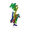
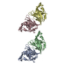
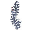
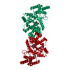
| Structure viewer | Molecule: MolmilJmol/JSmol |
|---|
- Downloads & links
-Download
| PDBx/mmCIF format | 5ws3.cif.gz | 224.6 KB | Display | PDBx/mmCIF format |
|---|---|---|---|---|
| PDB format | pdb5ws3.ent.gz | 179.3 KB | Display | PDB format |
| PDBx/mmJSON format | 5ws3.json.gz | Tree view | PDBx/mmJSON format | |
| Others |  Other downloads Other downloads |
-Validation report
| Arichive directory | https://data.pdbj.org/pub/pdb/validation_reports/ws/5ws3ftp://data.pdbj.org/pub/pdb/validation_reports/ws/5ws3 | HTTPS FTP |
|---|
-Related structure data
| Related structure data |  5wqcC  4s0vS S: Starting model for refinement C: citing same article ( |
|---|---|
| Similar structure data | |
| Experimental dataset #1 | Data reference: 10.11577/1413670 / Data set type: diffraction image data |


-Links
 PDBj
PDBj


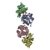
- Assembly
| Deposited unit | 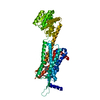
| ||||||||
|---|---|---|---|---|---|---|---|---|---|
| 1 |
| ||||||||
| Unit cell |
|
-Components
| #1: Protein | Mass: 64204.871 Da / Num. of mol.: 1 Fragment: UNP residues 3-254,UNP residues 218-413,UNP residues 294-388 Source method: isolated from a genetically manipulated source Details: Chimera protein of UNP residues 3-254 from Orexin receptor type 2 (O43614), UNP residues 218-413 from GlgA glycogen synthase (Q9V2J8), UNP residues 294-388 from Orexin receptor type 2 (O43614) Source: (gene. exp.) Homo sapiens (human), (gene. exp.) Pyrococcus abyssi (strain GE5 / Orsay) (archaea)Gene: HCRTR2, PAB2292 / Strain: GE5 / Orsay / Production host:   Spodoptera frugiperda (fall armyworm) / References: UniProt: O43614, UniProt: Q9V2J8 Spodoptera frugiperda (fall armyworm) / References: UniProt: O43614, UniProt: Q9V2J8 | ||||||
|---|---|---|---|---|---|---|---|
| #2: Chemical | ChemComp-7MA / 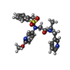  Mass: 454.542 Da / Num. of mol.: 1 / Source method: obtained synthetically / Formula: C23H26N4O4S / Comment: antagonist*YM Mass: 454.542 Da / Num. of mol.: 1 / Source method: obtained synthetically / Formula: C23H26N4O4S / Comment: antagonist*YM | ||||||
| #3: Chemical | ChemComp-OLA /   Mass: 282.461 Da / Num. of mol.: 4 / Source method: obtained synthetically / Formula: C18H34O2 Mass: 282.461 Da / Num. of mol.: 4 / Source method: obtained synthetically / Formula: C18H34O2#4: Chemical |   Mass: 238.278 Da / Num. of mol.: 2 / Source method: obtained synthetically / Formula: C10H22O6 / Comment: precipitant*YM Mass: 238.278 Da / Num. of mol.: 2 / Source method: obtained synthetically / Formula: C10H22O6 / Comment: precipitant*YM#5: Water | ChemComp-HOH / |  Mass: 18.015 Da / Num. of mol.: 12 / Source method: isolated from a natural source / Formula: H2O Mass: 18.015 Da / Num. of mol.: 12 / Source method: isolated from a natural source / Formula: H2OHas protein modification | Y | |
-Experimental details
-Experiment
| Experiment | Method: X-RAY DIFFRACTION / Number of used crystals: 1 |
|---|
- Sample preparation
| Crystal | Density Matthews: 2.53 Å3/Da / Density % sol: 51.42 % |
|---|---|
| Crystal grow | Temperature: 293 K / Method: lipidic cubic phase / pH: 6 Details: 0.1M MES, 0.1M sodium malonate, 27-30% PEG 300, pH 6.0 |
-Data collection
| Diffraction | Mean temperature: 293 K |
|---|---|
| Diffraction source | Source: FREE ELECTRON LASER / Site: SACLA / Beamline: BL3 / Wavelength: 1.77 Å |
| Detector | Type: MPCCD / Detector: CCD / Date: Apr 3, 2015 |
| Radiation | Protocol: SINGLE WAVELENGTH / Monochromatic (M) / Laue (L): M / Scattering type: x-ray |
| Radiation wavelength | Wavelength: 1.77 Å / Relative weight: 1 |
| Reflection | Resolution: 2.3→50 Å / Num. obs: 28958 / % possible obs: 100 % / Redundancy: 77.6 % / CC1/2: 0.98 / Net I/σ(I): 5.5 |
| Reflection shell | Resolution: 2.3→2.38 Å / Redundancy: 7.2 % / CC1/2: 0.58 / % possible all: 98.4 |
- Processing
| Software |
| |||||||||||||||||||||||||||||||||||||||||||||||||||||||||||||||||||||||||||||
|---|---|---|---|---|---|---|---|---|---|---|---|---|---|---|---|---|---|---|---|---|---|---|---|---|---|---|---|---|---|---|---|---|---|---|---|---|---|---|---|---|---|---|---|---|---|---|---|---|---|---|---|---|---|---|---|---|---|---|---|---|---|---|---|---|---|---|---|---|---|---|---|---|---|---|---|---|---|---|
| Refinement | Method to determine structure: MOLECULAR REPLACEMENT Starting model: 4s0v Resolution: 2.3→45.109 Å / SU ML: 0.37 / Cross valid method: FREE R-VALUE / σ(F): 1.34 / Phase error: 28.5 / Stereochemistry target values: ML
| |||||||||||||||||||||||||||||||||||||||||||||||||||||||||||||||||||||||||||||
| Solvent computation | Shrinkage radii: 0.9 Å / VDW probe radii: 1.11 Å / Solvent model: FLAT BULK SOLVENT MODEL | |||||||||||||||||||||||||||||||||||||||||||||||||||||||||||||||||||||||||||||
| Refinement step | Cycle: LAST / Resolution: 2.3→45.109 Å
| |||||||||||||||||||||||||||||||||||||||||||||||||||||||||||||||||||||||||||||
| Refine LS restraints |
| |||||||||||||||||||||||||||||||||||||||||||||||||||||||||||||||||||||||||||||
| LS refinement shell |
| |||||||||||||||||||||||||||||||||||||||||||||||||||||||||||||||||||||||||||||
| Refinement TLS params. | Method: refined / Refine-ID: X-RAY DIFFRACTION
| |||||||||||||||||||||||||||||||||||||||||||||||||||||||||||||||||||||||||||||
| Refinement TLS group |
|
