 Movie
Movie Controller

[English] 日本語
 Yorodumi
- PDB-5wob: Crystal Structure Analysis of Fab1-Bound Human Insulin Degrading ... -
+ Open data

- Basic information
| Entry | Database: PDB / ID: 5wob | |||||||||
|---|---|---|---|---|---|---|---|---|---|---|
| Title | Crystal Structure Analysis of Fab1-Bound Human Insulin Degrading Enzyme (IDE) in Complex with Insulin | |||||||||
 Components Components |
| |||||||||
 Keywords Keywords | HYDROLASE / complex | |||||||||
| Function / homology |  Function and homology information Function and homology informationinsulysin / beta-endorphin binding / ubiquitin recycling / insulin catabolic process / insulin metabolic process / amyloid-beta clearance by cellular catabolic process / hormone catabolic process / bradykinin catabolic process / cytosolic proteasome complex / positive regulation of protein binding ...insulysin / beta-endorphin binding / ubiquitin recycling / insulin catabolic process / insulin metabolic process / amyloid-beta clearance by cellular catabolic process / hormone catabolic process / bradykinin catabolic process / cytosolic proteasome complex / positive regulation of protein binding / insulin binding / negative regulation of glycogen catabolic process / : / regulation of aerobic respiration / negative regulation of fatty acid metabolic process / Signaling by Insulin receptor / negative regulation of feeding behavior / peptide catabolic process / IRS activation / Insulin processing / regulation of protein secretion / positive regulation of peptide hormone secretion / negative regulation of acute inflammatory response / Regulation of gene expression in beta cells / positive regulation of respiratory burst / peroxisomal matrix / alpha-beta T cell activation / amyloid-beta clearance / Synthesis, secretion, and deacylation of Ghrelin / amyloid-beta metabolic process / negative regulation of protein secretion / positive regulation of dendritic spine maintenance / negative regulation of gluconeogenesis / fatty acid homeostasis / positive regulation of glycogen biosynthetic process / positive regulation of insulin receptor signaling pathway / Signal attenuation / FOXO-mediated transcription of oxidative stress, metabolic and neuronal genes / negative regulation of lipid catabolic process / negative regulation of respiratory burst involved in inflammatory response / positive regulation of lipid biosynthetic process / negative regulation of oxidative stress-induced intrinsic apoptotic signaling pathway / nitric oxide-cGMP-mediated signaling / regulation of protein localization to plasma membrane / positive regulation of nitric-oxide synthase activity / transport vesicle / Insulin receptor recycling / COPI-mediated anterograde transport / positive regulation of brown fat cell differentiation / negative regulation of reactive oxygen species biosynthetic process / insulin-like growth factor receptor binding / negative regulation of proteolysis / NPAS4 regulates expression of target genes / peptide binding / neuron projection maintenance / positive regulation of mitotic nuclear division / endoplasmic reticulum-Golgi intermediate compartment membrane / Insulin receptor signalling cascade / : / positive regulation of glycolytic process / endosome lumen / positive regulation of cytokine production / acute-phase response / positive regulation of D-glucose import across plasma membrane / insulin receptor binding / protein catabolic process / positive regulation of long-term synaptic potentiation / positive regulation of protein secretion / positive regulation of cell differentiation / wound healing / Peroxisomal protein import / Regulation of insulin secretion / hormone activity / antigen processing and presentation of endogenous peptide antigen via MHC class I / positive regulation of neuron projection development / metalloendopeptidase activity / negative regulation of protein catabolic process / regulation of synaptic plasticity / positive regulation of protein localization to nucleus / Golgi lumen / cognition / glucose metabolic process / vasodilation / positive regulation of protein catabolic process / insulin receptor signaling pathway / peroxisome / cell-cell signaling / regulation of protein localization / glucose homeostasis / amyloid-beta binding / PI5P, PP2A and IER3 Regulate PI3K/AKT Signaling / virus receptor activity / positive regulation of cell growth / protease binding / secretory granule lumen / endopeptidase activity / basolateral plasma membrane / positive regulation of MAPK cascade / positive regulation of canonical NF-kappaB signal transduction / positive regulation of phosphatidylinositol 3-kinase/protein kinase B signal transduction Similarity search - Function | |||||||||
| Biological species |  Homo sapiens (human) Homo sapiens (human) | |||||||||
| Method |  X-RAY DIFFRACTION / SYNCHROTRON / Resolution: 3.95 Å X-RAY DIFFRACTION / SYNCHROTRON / Resolution: 3.95 Å | |||||||||
 Authors Authors | McCord, L.A. / Liang, W.G. / Farcasanu, M. / Wang, A.G. / Koide, S. / Tang, W.J. | |||||||||
| Funding support |  United States, 1items United States, 1items
| |||||||||
 Citation Citation | Journal: Elife / Year: 2018 Title: Ensemble cryoEM elucidates the mechanism of insulin capture and degradation by human insulin degrading enzyme. Authors: Zhening Zhang / Wenguang G Liang / Lucas J Bailey / Yong Zi Tan / Hui Wei / Andrew Wang / Mara Farcasanu / Virgil A Woods / Lauren A McCord / David Lee / Weifeng Shang / Rebecca Deprez- ...Authors: Zhening Zhang / Wenguang G Liang / Lucas J Bailey / Yong Zi Tan / Hui Wei / Andrew Wang / Mara Farcasanu / Virgil A Woods / Lauren A McCord / David Lee / Weifeng Shang / Rebecca Deprez-Poulain / Benoit Deprez / David R Liu / Akiko Koide / Shohei Koide / Anthony A Kossiakoff / Sheng Li / Bridget Carragher / Clinton S Potter / Wei-Jen Tang /  Abstract: Insulin degrading enzyme (IDE) plays key roles in degrading peptides vital in type two diabetes, Alzheimer's, inflammation, and other human diseases. However, the process through which IDE recognizes ...Insulin degrading enzyme (IDE) plays key roles in degrading peptides vital in type two diabetes, Alzheimer's, inflammation, and other human diseases. However, the process through which IDE recognizes peptides that tend to form amyloid fibrils remained unsolved. We used cryoEM to understand both the apo- and insulin-bound dimeric IDE states, revealing that IDE displays a large opening between the homologous ~55 kDa N- and C-terminal halves to allow selective substrate capture based on size and charge complementarity. We also used cryoEM, X-ray crystallography, SAXS, and HDX-MS to elucidate the molecular basis of how amyloidogenic peptides stabilize the disordered IDE catalytic cleft, thereby inducing selective degradation by substrate-assisted catalysis. Furthermore, our insulin-bound IDE structures explain how IDE processively degrades insulin by stochastically cutting either chain without breaking disulfide bonds. Together, our studies provide a mechanism for how IDE selectively degrades amyloidogenic peptides and offers structural insights for developing IDE-based therapies. | |||||||||
| History |
|
- Structure visualization
| Structure viewer | Molecule: MolmilJmol/JSmol |
|---|
- Downloads & links
-Download
| PDBx/mmCIF format | 5wob.cif.gz | 2.1 MB | Display | PDBx/mmCIF format |
|---|---|---|---|---|
| PDB format | pdb5wob.ent.gz | 1.7 MB | Display | PDB format |
| PDBx/mmJSON format | 5wob.json.gz | Tree view | PDBx/mmJSON format | |
| Others |  Other downloads Other downloads |
-Validation report
| Arichive directory | https://data.pdbj.org/pub/pdb/validation_reports/wo/5wobftp://data.pdbj.org/pub/pdb/validation_reports/wo/5wob | HTTPS FTP |
|---|
-Related structure data
| Related structure data | 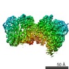 7041C 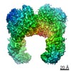 7062C 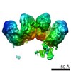 7065C  7066C 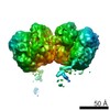 7090C 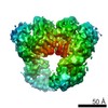 7091C 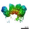 7092C 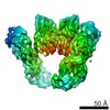 7093C 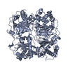 6b3qC 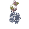 6b70C 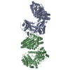 6b7yC 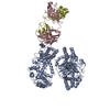 6b7zC 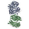 6bf6C 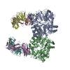 6bf7C 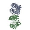 6bf8C 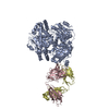 6bf9C 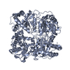 6bfcC 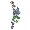 4iofS S: Starting model for refinement C: citing same article ( |
|---|---|
| Similar structure data |


-Links
 PDBj
PDBj


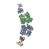
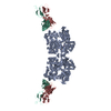
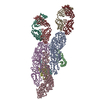
- Assembly
| Deposited unit | 
| ||||||||
|---|---|---|---|---|---|---|---|---|---|
| 1 | 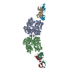
| ||||||||
| 2 | 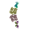
| ||||||||
| 3 | 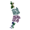
| ||||||||
| 4 | 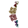
| ||||||||
| Unit cell |
|
-Components
| #1: Protein | Mass: 114560.578 Da / Num. of mol.: 8 / Fragment: UNP residues 42-1019 Source method: isolated from a genetically manipulated source Source: (gene. exp.) Homo sapiens (human) / Gene: IDE / Production host:  #2: Protein/peptide | Mass: 2269.595 Da / Num. of mol.: 8 Source method: isolated from a genetically manipulated source Source: (gene. exp.) Homo sapiens (human) / Gene: INS / Production host:  #3: Antibody | Mass: 28201.670 Da / Num. of mol.: 8 Source method: isolated from a genetically manipulated source Source: (gene. exp.) #4: Antibody | Mass: 25982.098 Da / Num. of mol.: 8 Source method: isolated from a genetically manipulated source Source: (gene. exp.) #5: Chemical | ChemComp-ZN /   Mass: 65.409 Da / Num. of mol.: 8 / Source method: obtained synthetically / Formula: Zn / Feature type: SUBJECT OF INVESTIGATION Mass: 65.409 Da / Num. of mol.: 8 / Source method: obtained synthetically / Formula: Zn / Feature type: SUBJECT OF INVESTIGATIONHas protein modification | Y | |
|---|
-Experimental details
-Experiment
| Experiment | Method: X-RAY DIFFRACTION / Number of used crystals: 1 |
|---|
- Sample preparation
| Crystal | Density Matthews: 2.28 Å3/Da / Density % sol: 46.08 % |
|---|---|
| Crystal grow | Temperature: 291.15 K / Method: vapor diffusion, hanging drop / pH: 6.5 Details: 0.1M Sodium cacodylate, pH6.5; 0.2M MgCl2; 10% PEG3000, VAPOR DIFFUSION, HANGING DROP, temperature 291.15K |
-Data collection
| Diffraction | Mean temperature: 100 K |
|---|---|
| Diffraction source | Source: SYNCHROTRON / Site: APS / Beamline: 19-ID / Wavelength: 0.9792 Å |
| Detector | Type: ADSC QUANTUM 315r / Detector: CCD / Date: Jul 11, 2013 |
| Radiation | Protocol: SINGLE WAVELENGTH / Monochromatic (M) / Laue (L): M / Scattering type: x-ray |
| Radiation wavelength | Wavelength: 0.9792 Å / Relative weight: 1 |
| Reflection | Resolution: 3.95→50 Å / Num. obs: 108370 / % possible obs: 99.5 % / Observed criterion σ(F): 2.1 / Observed criterion σ(I): 2.1 / Redundancy: 3.3 % / Rmerge(I) obs: 0.2 / Rpim(I) all: 0.13 / Rsym value: 0.12 / Χ2: 1.277 / Net I/σ(I): 7 |
| Reflection shell | Resolution: 3.95→4.02 Å / Redundancy: 3.3 % / Rmerge(I) obs: 0.672 / Mean I/σ(I) obs: 2.1 / Num. unique obs: 5406 / CC1/2: 0.583 / Rpim(I) all: 0.424 / Rsym value: 0.621 / Χ2: 1.02 / % possible all: 99.8 |
- Processing
| Software |
| |||||||||||||||||||||||||||||||||||||||||||||||||||||||||||||||||||||||||||||||||||||||||||||||||||||||||
|---|---|---|---|---|---|---|---|---|---|---|---|---|---|---|---|---|---|---|---|---|---|---|---|---|---|---|---|---|---|---|---|---|---|---|---|---|---|---|---|---|---|---|---|---|---|---|---|---|---|---|---|---|---|---|---|---|---|---|---|---|---|---|---|---|---|---|---|---|---|---|---|---|---|---|---|---|---|---|---|---|---|---|---|---|---|---|---|---|---|---|---|---|---|---|---|---|---|---|---|---|---|---|---|---|---|---|
| Refinement | Starting model: 4IOF Resolution: 3.95→49.543 Å / Cross valid method: FREE R-VALUE / σ(F): 1.34 / Phase error: 26.49
| |||||||||||||||||||||||||||||||||||||||||||||||||||||||||||||||||||||||||||||||||||||||||||||||||||||||||
| Solvent computation | Shrinkage radii: 0.9 Å / VDW probe radii: 1.11 Å | |||||||||||||||||||||||||||||||||||||||||||||||||||||||||||||||||||||||||||||||||||||||||||||||||||||||||
| Refinement step | Cycle: LAST / Resolution: 3.95→49.543 Å
| |||||||||||||||||||||||||||||||||||||||||||||||||||||||||||||||||||||||||||||||||||||||||||||||||||||||||
| Refine LS restraints |
| |||||||||||||||||||||||||||||||||||||||||||||||||||||||||||||||||||||||||||||||||||||||||||||||||||||||||
| LS refinement shell |
|
