 Movie
Movie Controller

[English] 日本語
 Yorodumi
- PDB-4m1c: Crystal Structure Analysis of Fab-Bound Human Insulin Degrading E... -
+ Open data

- Basic information
| Entry | Database: PDB / ID: 4m1c | ||||||
|---|---|---|---|---|---|---|---|
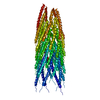
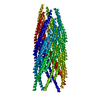
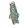
| Title | Crystal Structure Analysis of Fab-Bound Human Insulin Degrading Enzyme (IDE) in Complex with Amyloid-Beta (1-40) | ||||||
 Components Components |
| ||||||
 Keywords Keywords | HYDROLASE / Zinc metalloprotease | ||||||
| Function / homology |  Function and homology information Function and homology informationinsulysin / beta-endorphin binding / ubiquitin recycling / insulin catabolic process / insulin metabolic process / amyloid-beta clearance by cellular catabolic process / hormone catabolic process / bradykinin catabolic process / amyloid-beta complex / growth cone lamellipodium ...insulysin / beta-endorphin binding / ubiquitin recycling / insulin catabolic process / insulin metabolic process / amyloid-beta clearance by cellular catabolic process / hormone catabolic process / bradykinin catabolic process / amyloid-beta complex / growth cone lamellipodium / cellular response to norepinephrine stimulus / collateral sprouting in absence of injury / growth cone filopodium / microglia development / Formyl peptide receptors bind formyl peptides and many other ligands / regulation of Wnt signaling pathway / axo-dendritic transport / cytosolic proteasome complex / axon midline choice point recognition / hippocampal neuron apoptotic process / regulation of synapse structure or activity / astrocyte activation involved in immune response / NMDA selective glutamate receptor signaling pathway / positive regulation of protein binding / regulation of spontaneous synaptic transmission / mating behavior / growth factor receptor binding / insulin binding / peptidase activator activity / regulation of aerobic respiration / Insertion of tail-anchored proteins into the endoplasmic reticulum membrane / positive regulation of amyloid fibril formation / Golgi-associated vesicle / PTB domain binding / peptide catabolic process / astrocyte projection / Lysosome Vesicle Biogenesis / Deregulated CDK5 triggers multiple neurodegenerative pathways in Alzheimer's disease models / neuron remodeling / nuclear envelope lumen / TRAF6 mediated NF-kB activation / dendrite development / peroxisomal matrix / positive regulation of protein metabolic process / signaling receptor activator activity / amyloid-beta clearance / negative regulation of long-term synaptic potentiation / Advanced glycosylation endproduct receptor signaling / transition metal ion binding / The NLRP3 inflammasome / amyloid-beta metabolic process / modulation of excitatory postsynaptic potential / regulation of multicellular organism growth / main axon / intracellular copper ion homeostasis / ECM proteoglycans / response to insulin-like growth factor stimulus / positive regulation of T cell migration / regulation of presynapse assembly / neuronal dense core vesicle / Purinergic signaling in leishmaniasis infection / Insulin receptor recycling / cellular response to manganese ion / Notch signaling pathway / negative regulation of proteolysis / positive regulation of chemokine production / swimming behavior / peptide binding / neuron projection maintenance / extracellular matrix organization / clathrin-coated pit / positive regulation of mitotic cell cycle / axonogenesis / Mitochondrial protein degradation / positive regulation of calcium-mediated signaling / ionotropic glutamate receptor signaling pathway / platelet alpha granule lumen / astrocyte activation / response to interleukin-1 / : / cellular response to cAMP / regulation of neuron apoptotic process / cellular response to copper ion / positive regulation of glycolytic process / endosome lumen / trans-Golgi network membrane / positive regulation of interleukin-1 beta production / protein catabolic process / protein serine/threonine kinase binding / dendritic shaft / positive regulation of long-term synaptic potentiation / learning / central nervous system development / Post-translational protein phosphorylation / adult locomotory behavior / serine-type endopeptidase inhibitor activity / Peroxisomal protein import / locomotory behavior / microglial cell activation / antigen processing and presentation of endogenous peptide antigen via MHC class I Similarity search - Function | ||||||
| Biological species |  Homo sapiens (human) Homo sapiens (human) | ||||||
| Method |  X-RAY DIFFRACTION / SYNCHROTRON / MOLECULAR REPLACEMENT / Resolution: 3.5007 Å X-RAY DIFFRACTION / SYNCHROTRON / MOLECULAR REPLACEMENT / Resolution: 3.5007 Å | ||||||
 Authors Authors | McCord, L.M. / Liang, W. / Farcasanu, M. / Scherpelz, K. / Meredith, S.C. / Koide, S. / Tang, W.J. | ||||||
 Citation Citation | Journal: To be Published Title: Crystal Structure Analysis of Fab-Bound Human Insulin Degrading Enzyme (IDE) in Complex with Amyloid-Beta (1-40) Authors: McCord, L.A. / Liang, W. / Farcasanu, M. / Scherpelz, K. / Meredith, S.C. / Koide, S. / Tang, W.J. | ||||||
| History |
|
- Structure visualization
| Structure viewer | Molecule: MolmilJmol/JSmol |
|---|
- Downloads & links
-Download
| PDBx/mmCIF format | 4m1c.cif.gz | 1.1 MB | Display | PDBx/mmCIF format |
|---|---|---|---|---|
| PDB format | pdb4m1c.ent.gz | 889.5 KB | Display | PDB format |
| PDBx/mmJSON format | 4m1c.json.gz | Tree view | PDBx/mmJSON format | |
| Others |  Other downloads Other downloads |
-Validation report
| Arichive directory | https://data.pdbj.org/pub/pdb/validation_reports/m1/4m1cftp://data.pdbj.org/pub/pdb/validation_reports/m1/4m1c | HTTPS FTP |
|---|
-Related structure data
| Related structure data | 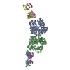 4iofS S: Starting model for refinement |
|---|---|
| Similar structure data |


-Links
 PDBj
PDBj


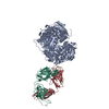
- Assembly
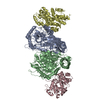
| Deposited unit | 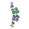
| |||||||||||||||
|---|---|---|---|---|---|---|---|---|---|---|---|---|---|---|---|---|
| 1 | 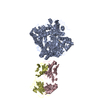
| |||||||||||||||
| 2 | 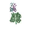
| |||||||||||||||
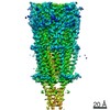
| Unit cell |
| |||||||||||||||
| Noncrystallographic symmetry (NCS) | NCS domain:
|
-Components
| #1: Protein | Mass: 114560.578 Da / Num. of mol.: 2 / Mutation: E111Q Source method: isolated from a genetically manipulated source Details: Cysteine-Free / Source: (gene. exp.) Homo sapiens (human) / Gene: IDE, Insulin Degrading Enzyme / Production host:  #2: Antibody | Mass: 28201.670 Da / Num. of mol.: 2 / Source method: obtained synthetically Details: Specific antigen-binding fragment (Fab, light chain) synthetically engineered to target Human Insulin Degrading Enzyme Source: (synth.) Homo sapiens (human)#3: Antibody | Mass: 25982.098 Da / Num. of mol.: 2 / Source method: obtained synthetically Details: Specific antigen-binding fragment (Fab, heavy chain) synthetically engineered to target Human Insulin Degrading Enzyme Source: (synth.) Homo sapiens (human)#4: Protein/peptide | Mass: 4335.852 Da / Num. of mol.: 2 / Source method: obtained synthetically / Details: Amyloid-Beta (1-40) / Source: (synth.) Homo sapiens (human) / References: UniProt: P05067#5: Chemical |   Mass: 65.409 Da / Num. of mol.: 2 / Source method: obtained synthetically / Formula: Zn Mass: 65.409 Da / Num. of mol.: 2 / Source method: obtained synthetically / Formula: ZnHas protein modification | Y | |
|---|
-Experimental details
-Experiment
| Experiment | Method: X-RAY DIFFRACTION / Number of used crystals: 1 |
|---|
- Sample preparation
| Crystal | Density Matthews: 2.04 Å3/Da / Density % sol: 39.65 % |
|---|---|
| Crystal grow | Temperature: 291.15 K / Method: vapor diffusion, hanging drop / pH: 6.5 Details: 0.1M Sodium cacodylate, pH6.5, 0.2M MgCl2, 10% PEG-3000, VAPOR DIFFUSION, HANGING DROP, temperature 291.15K |
-Data collection
| Diffraction source | Source: SYNCHROTRON / Site: APS  / Beamline: 19-ID / Wavelength: 0.9792 Å / Beamline: 19-ID / Wavelength: 0.9792 Å |
|---|---|
| Detector | Type: ADSC QUANTUM 315r / Detector: CCD / Date: Jun 10, 2013 |
| Radiation | Monochromator: MAR CCD / Protocol: SINGLE WAVELENGTH / Monochromatic (M) / Laue (L): M / Scattering type: x-ray |
| Radiation wavelength | Wavelength: 0.9792 Å / Relative weight: 1 |
| Reflection | Resolution: 3.5→49.1 Å / Num. all: 35563 / Num. obs: 35563 / % possible obs: 96.6 % / Redundancy: 5.5 % / Biso Wilson estimate: 52.45 Å2 / Rmerge(I) obs: 0.159 |
| Reflection shell | Resolution: 3.5→3.56 Å / Redundancy: 5.6 % / Rmerge(I) obs: 0.68 / % possible all: 96.7 |
- Processing
| Software |
| |||||||||||||||||||||||||||||||||||||||||||||||||||||||||||||||||||||||||||||||||||||||||||||||||||||||||
|---|---|---|---|---|---|---|---|---|---|---|---|---|---|---|---|---|---|---|---|---|---|---|---|---|---|---|---|---|---|---|---|---|---|---|---|---|---|---|---|---|---|---|---|---|---|---|---|---|---|---|---|---|---|---|---|---|---|---|---|---|---|---|---|---|---|---|---|---|---|---|---|---|---|---|---|---|---|---|---|---|---|---|---|---|---|---|---|---|---|---|---|---|---|---|---|---|---|---|---|---|---|---|---|---|---|---|
| Refinement | Method to determine structure: MOLECULAR REPLACEMENT Starting model: PDB ENTRY 4IOF Resolution: 3.5007→49.064 Å / SU ML: 0.45 / σ(F): 1.34 / Phase error: 26.98 / Stereochemistry target values: ML
| |||||||||||||||||||||||||||||||||||||||||||||||||||||||||||||||||||||||||||||||||||||||||||||||||||||||||
| Solvent computation | Shrinkage radii: 0.9 Å / VDW probe radii: 1.11 Å / Solvent model: FLAT BULK SOLVENT MODEL | |||||||||||||||||||||||||||||||||||||||||||||||||||||||||||||||||||||||||||||||||||||||||||||||||||||||||
| Refinement step | Cycle: LAST / Resolution: 3.5007→49.064 Å
| |||||||||||||||||||||||||||||||||||||||||||||||||||||||||||||||||||||||||||||||||||||||||||||||||||||||||
| Refine LS restraints |
| |||||||||||||||||||||||||||||||||||||||||||||||||||||||||||||||||||||||||||||||||||||||||||||||||||||||||
| LS refinement shell |
| |||||||||||||||||||||||||||||||||||||||||||||||||||||||||||||||||||||||||||||||||||||||||||||||||||||||||
| Refinement TLS params. | Method: refined / Origin x: 13.9556 Å / Origin y: -8.5682 Å / Origin z: -98.8944 Å
| |||||||||||||||||||||||||||||||||||||||||||||||||||||||||||||||||||||||||||||||||||||||||||||||||||||||||
| Refinement TLS group | Selection details: all |
