 Movie
Movie Controller

[English] 日本語
 Yorodumi
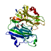
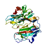
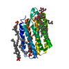
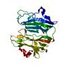
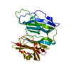
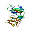
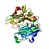
- PDB-5wja: Crystal structure of H107A peptidylglycine alpha-hydroxylating mo... -
+ Open data

- Basic information
| Entry | Database: PDB / ID: 5wja | ||||||
|---|---|---|---|---|---|---|---|
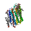
| Title | Crystal structure of H107A peptidylglycine alpha-hydroxylating monooxygenase (PHM) in complex with citrate | ||||||
 Components Components | Peptidyl-glycine alpha-amidating monooxygenase | ||||||
 Keywords Keywords | OXIDOREDUCTASE / PEPTIDYLGLYCINE MONOOXYGENASE / PEPTIDYLGLYCINE 2-HYDROXYLASE / PHM / METAL BINDING PROTEIN / LYASE | ||||||
| Function / homology |  Function and homology information Function and homology informationpeptidylglycine monooxygenase / peptidylamidoglycolate lyase / peptide amidation / peptidylglycine monooxygenase activity / peptidylamidoglycolate lyase activity / fatty acid primary amide biosynthetic process / ovulation cycle process / toxin metabolic process / long-chain fatty acid metabolic process / peptide metabolic process ...peptidylglycine monooxygenase / peptidylamidoglycolate lyase / peptide amidation / peptidylglycine monooxygenase activity / peptidylamidoglycolate lyase activity / fatty acid primary amide biosynthetic process / ovulation cycle process / toxin metabolic process / long-chain fatty acid metabolic process / peptide metabolic process / mitotic chromosome condensation / response to pH / L-ascorbic acid binding / response to zinc ion / response to copper ion / limb development / transport vesicle membrane / maternal process involved in female pregnancy / condensed chromosome / lactation / secretory granule / response to glucocorticoid / regulation of actin cytoskeleton organization / trans-Golgi network / response to estradiol / heart development / cellular response to oxidative stress / proteasome-mediated ubiquitin-dependent protein catabolic process / perikaryon / response to hypoxia / response to xenobiotic stimulus / copper ion binding / neuronal cell body / calcium ion binding / chromatin binding / regulation of transcription by RNA polymerase II / protein kinase binding / chromatin / perinuclear region of cytoplasm / cell surface / : / extracellular region / zinc ion binding / identical protein binding Similarity search - Function | ||||||
| Biological species |  | ||||||
| Method |  X-RAY DIFFRACTION / MOLECULAR REPLACEMENT / Resolution: 2.3 Å X-RAY DIFFRACTION / MOLECULAR REPLACEMENT / Resolution: 2.3 Å | ||||||
| Model details | copper M is present in both molecules: copper H is present in only one molecule | ||||||
 Authors Authors | Maheshwari, S. / Rudzka, K. / Gabelli, S.B. / Amzel, L.M. | ||||||
| Funding support |  United States, 1items United States, 1items
| ||||||
 Citation Citation | Journal: Commun Biol / Year: 2018 Title: Effects of copper occupancy on the conformational landscape of peptidylglycine alpha-hydroxylating monooxygenase. Authors: Maheshwari, S. / Shimokawa, C. / Rudzka, K. / Kline, C.D. / Eipper, B.A. / Mains, R.E. / Gabelli, S.B. / Blackburn, N. / Amzel, L.M. | ||||||
| History |
|
- Structure visualization
| Structure viewer | Molecule: MolmilJmol/JSmol |
|---|
- Downloads & links
-Download
| PDBx/mmCIF format | 5wja.cif.gz | 139.7 KB | Display | PDBx/mmCIF format |
|---|---|---|---|---|
| PDB format | pdb5wja.ent.gz | 106.8 KB | Display | PDB format |
| PDBx/mmJSON format | 5wja.json.gz | Tree view | PDBx/mmJSON format | |
| Others |  Other downloads Other downloads |
-Validation report
| Arichive directory | https://data.pdbj.org/pub/pdb/validation_reports/wj/5wjaftp://data.pdbj.org/pub/pdb/validation_reports/wj/5wja | HTTPS FTP |
|---|
-Related structure data
| Related structure data | 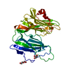 5wkwC 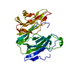 5wm0C 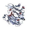 6alaC 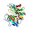 6alvC 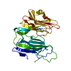 6ampC 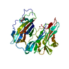 6an3C 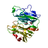 6ao6C 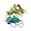 6ay0C 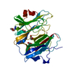 1phmS S: Starting model for refinement C: citing same article ( |
|---|---|
| Similar structure data |


-Links
 PDBj
PDBj


- Assembly
| Deposited unit | 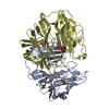
| ||||||||
|---|---|---|---|---|---|---|---|---|---|
| 1 | 
| ||||||||
| 2 | 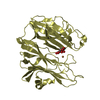
| ||||||||
| Unit cell |
|
-Components
-Protein , 1 types, 2 molecules AD
| #1: Protein | Mass: 34706.812 Da / Num. of mol.: 2 / Fragment: PHM / Mutation: H107A Source method: isolated from a genetically manipulated source Source: (gene. exp.)  Cricetulus griseus (Chinese hamster) / Strain (production host): CHO Cricetulus griseus (Chinese hamster) / Strain (production host): CHOReferences: UniProt: P14925, peptidylglycine monooxygenase, peptidylamidoglycolate lyase |
|---|
-Non-polymers , 5 types, 153 molecules 


| #2: Chemical |  Mass: 63.546 Da / Num. of mol.: 3 / Source method: obtained synthetically / Formula: Cu Mass: 63.546 Da / Num. of mol.: 3 / Source method: obtained synthetically / Formula: Cu#3: Chemical | ChemComp-GOL /  Mass: 92.094 Da / Num. of mol.: 6 / Source method: obtained synthetically / Formula: C3H8O3 Mass: 92.094 Da / Num. of mol.: 6 / Source method: obtained synthetically / Formula: C3H8O3#4: Chemical | ChemComp-NI / |  Mass: 58.693 Da / Num. of mol.: 1 / Source method: obtained synthetically / Formula: Ni Mass: 58.693 Da / Num. of mol.: 1 / Source method: obtained synthetically / Formula: Ni#5: Chemical | ChemComp-FLC / |  Mass: 189.100 Da / Num. of mol.: 1 / Source method: obtained synthetically / Formula: C6H5O7 Mass: 189.100 Da / Num. of mol.: 1 / Source method: obtained synthetically / Formula: C6H5O7#6: Water | ChemComp-HOH / | Mass: 18.015 Da / Num. of mol.: 142 / Source method: isolated from a natural source / Formula: H2O |
|---|
-Details
| Has protein modification | Y |
|---|
-Experimental details
-Experiment
| Experiment | Method: X-RAY DIFFRACTION / Number of used crystals: 1 |
|---|
- Sample preparation
| Crystal | Density Matthews: 2.19 Å3/Da / Density % sol: 43.88 % / Mosaicity: 0.72 ° |
|---|---|
| Crystal grow | Temperature: 293 K / Method: vapor diffusion / pH: 8.5 / Details: 19-24% PEG 4000, Tris HCL, citrate |
-Data collection
| Diffraction | Mean temperature: 100 K | ||||||||||||||||||||||||||||||
|---|---|---|---|---|---|---|---|---|---|---|---|---|---|---|---|---|---|---|---|---|---|---|---|---|---|---|---|---|---|---|---|
| Diffraction source | Source: ROTATING ANODE / Type: RIGAKU FR-E DW / Wavelength: 1.5418 Å | ||||||||||||||||||||||||||||||
| Detector | Type: RIGAKU SATURN 944+ / Detector: CCD / Date: Jul 22, 2014 | ||||||||||||||||||||||||||||||
| Radiation | Protocol: SINGLE WAVELENGTH / Monochromatic (M) / Laue (L): M / Scattering type: x-ray | ||||||||||||||||||||||||||||||
| Radiation wavelength | Wavelength: 1.5418 Å / Relative weight: 1 | ||||||||||||||||||||||||||||||
| Reflection | Resolution: 2.3→29.77 Å / Num. obs: 27694 / % possible obs: 99.6 % / Redundancy: 1.9 % / CC1/2: 0.996 / Rmerge(I) obs: 0.048 / Rpim(I) all: 0.048 / Rrim(I) all: 0.069 / Net I/σ(I): 9.6 / Num. measured all: 52974 / Scaling rejects: 0 | ||||||||||||||||||||||||||||||
| Reflection shell | Diffraction-ID: 1
|
- Processing
| Software |
| ||||||||||||||||||||||||||||||||||||||||||||||||||||||||||||
|---|---|---|---|---|---|---|---|---|---|---|---|---|---|---|---|---|---|---|---|---|---|---|---|---|---|---|---|---|---|---|---|---|---|---|---|---|---|---|---|---|---|---|---|---|---|---|---|---|---|---|---|---|---|---|---|---|---|---|---|---|---|
| Refinement | Method to determine structure: MOLECULAR REPLACEMENT Starting model: 1PHM Resolution: 2.3→29.77 Å / Cor.coef. Fo:Fc: 0.956 / Cor.coef. Fo:Fc free: 0.922 / WRfactor Rfree: 0.2799 / WRfactor Rwork: 0.201 / FOM work R set: 0.7566 / SU B: 10.497 / SU ML: 0.244 / SU R Cruickshank DPI: 0.4148 / SU Rfree: 0.2795 / Cross valid method: THROUGHOUT / σ(F): 0 / ESU R: 0.415 / ESU R Free: 0.279 / Stereochemistry target values: MAXIMUM LIKELIHOOD Details: HYDROGENS HAVE BEEN ADDED IN THE RIDING POSITIONS U VALUES : REFINED INDIVIDUALLY
| ||||||||||||||||||||||||||||||||||||||||||||||||||||||||||||
| Solvent computation | Ion probe radii: 0.8 Å / Shrinkage radii: 0.8 Å / VDW probe radii: 1.2 Å / Solvent model: MASK | ||||||||||||||||||||||||||||||||||||||||||||||||||||||||||||
| Displacement parameters | Biso max: 148.68 Å2 / Biso mean: 51.392 Å2 / Biso min: 17.72 Å2
| ||||||||||||||||||||||||||||||||||||||||||||||||||||||||||||
| Refinement step | Cycle: final / Resolution: 2.3→29.77 Å
| ||||||||||||||||||||||||||||||||||||||||||||||||||||||||||||
| Refine LS restraints |
| ||||||||||||||||||||||||||||||||||||||||||||||||||||||||||||
| LS refinement shell | Resolution: 2.301→2.361 Å / Rfactor Rfree error: 0 / Total num. of bins used: 20
|
