 Movie
Movie Controller


+ Open data

- Basic information
| Entry | Database: PDB / ID: 5w3s | |||||||||||||||||||||||||||||||||||||||||||||||||||||||||||||||||||||||||||||||||||||||||||||||||||||||||||||||
|---|---|---|---|---|---|---|---|---|---|---|---|---|---|---|---|---|---|---|---|---|---|---|---|---|---|---|---|---|---|---|---|---|---|---|---|---|---|---|---|---|---|---|---|---|---|---|---|---|---|---|---|---|---|---|---|---|---|---|---|---|---|---|---|---|---|---|---|---|---|---|---|---|---|---|---|---|---|---|---|---|---|---|---|---|---|---|---|---|---|---|---|---|---|---|---|---|---|---|---|---|---|---|---|---|---|---|---|---|---|---|---|---|
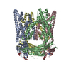
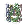
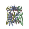
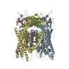
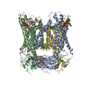
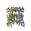
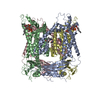
| Title | Cryo-electron microscopy structure of a TRPML3 ion channel | |||||||||||||||||||||||||||||||||||||||||||||||||||||||||||||||||||||||||||||||||||||||||||||||||||||||||||||||
 Components Components | Mucolipin-3 isoform 1 | |||||||||||||||||||||||||||||||||||||||||||||||||||||||||||||||||||||||||||||||||||||||||||||||||||||||||||||||
 Keywords Keywords | TRANSPORT PROTEIN / transient receptor potential channel / mucolipin / ion channel / membrane transport / TRPML / TRP channel / calcium channel / PIP2 / PI(3 / 5)P2 / lipid-gated channel / mucolipidosis / lysosomal ion channel / lysosome | |||||||||||||||||||||||||||||||||||||||||||||||||||||||||||||||||||||||||||||||||||||||||||||||||||||||||||||||
| Function / homology |  Function and homology information Function and homology informationstereocilium membrane / intracellularly phosphatidylinositol-3,5-bisphosphate-gated monatomic cation channel activity / NAADP-sensitive calcium-release channel activity / monoatomic anion channel activity / phosphatidylinositol-3,5-bisphosphate binding / sodium channel activity / autophagosome membrane / monoatomic cation transmembrane transport / potassium channel activity / late endosome membrane ...stereocilium membrane / intracellularly phosphatidylinositol-3,5-bisphosphate-gated monatomic cation channel activity / NAADP-sensitive calcium-release channel activity / monoatomic anion channel activity / phosphatidylinositol-3,5-bisphosphate binding / sodium channel activity / autophagosome membrane / monoatomic cation transmembrane transport / potassium channel activity / late endosome membrane / early endosome membrane / protein homotetramerization / lysosomal membrane / membrane / identical protein binding Similarity search - Function | |||||||||||||||||||||||||||||||||||||||||||||||||||||||||||||||||||||||||||||||||||||||||||||||||||||||||||||||
| Biological species |  | |||||||||||||||||||||||||||||||||||||||||||||||||||||||||||||||||||||||||||||||||||||||||||||||||||||||||||||||
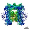
| Method | ELECTRON MICROSCOPY / single particle reconstruction / cryo EM / Resolution: 2.94 Å | |||||||||||||||||||||||||||||||||||||||||||||||||||||||||||||||||||||||||||||||||||||||||||||||||||||||||||||||
 Authors Authors | Hirschi, M. / Herzik, M.A. / Wie, J. / Suo, Y. / Borschel, W.F. / Ren, D. / Lander, G.C. / Lee, S.Y. | |||||||||||||||||||||||||||||||||||||||||||||||||||||||||||||||||||||||||||||||||||||||||||||||||||||||||||||||
| Funding support |  United States, 2items United States, 2items
| |||||||||||||||||||||||||||||||||||||||||||||||||||||||||||||||||||||||||||||||||||||||||||||||||||||||||||||||
 Citation Citation | Journal: Nature / Year: 2017 Title: Cryo-electron microscopy structure of the lysosomal calcium-permeable channel TRPML3. Authors: Marscha Hirschi / Mark A Herzik / Jinhong Wie / Yang Suo / William F Borschel / Dejian Ren / Gabriel C Lander / Seok-Yong Lee / Abstract: The modulation of ion channel activity by lipids is increasingly recognized as a fundamental component of cellular signalling. The transient receptor potential mucolipin (TRPML) channel family ...The modulation of ion channel activity by lipids is increasingly recognized as a fundamental component of cellular signalling. The transient receptor potential mucolipin (TRPML) channel family belongs to the TRP superfamily and is composed of three members: TRPML1-TRPML3. TRPMLs are the major Ca-permeable channels on late endosomes and lysosomes (LEL). They regulate the release of Ca from organelles, which is important for various physiological processes, including organelle trafficking and fusion. Loss-of-function mutations in the MCOLN1 gene, which encodes TRPML1, cause the neurodegenerative lysosomal storage disorder mucolipidosis type IV, and a gain-of-function mutation (Ala419Pro) in TRPML3 gives rise to the varitint-waddler (Va) mouse phenotype. Notably, TRPML channels are activated by the low-abundance and LEL-enriched signalling lipid phosphatidylinositol-3,5-bisphosphate (PtdIns(3,5)P), whereas other phosphoinositides such as PtdIns(4,5)P, which is enriched in plasma membranes, inhibit TRPMLs. Conserved basic residues at the N terminus of the channel are important for activation by PtdIns(3,5)P and inhibition by PtdIns(4,5)P. However, owing to a lack of structural information, the mechanism by which TRPML channels recognize PtdIns(3,5)P and increase their Ca conductance remains unclear. Here we present the cryo-electron microscopy (cryo-EM) structure of a full-length TRPML3 channel from the common marmoset (Callithrix jacchus) at an overall resolution of 2.9 Å. Our structure reveals not only the molecular basis of ion conduction but also the unique architecture of TRPMLs, wherein the voltage sensor-like domain is linked to the pore via a cytosolic domain that we term the mucolipin domain. Combined with functional studies, these data suggest that the mucolipin domain is responsible for PtdIns(3,5)P binding and subsequent channel activation, and that it acts as a 'gating pulley' for lipid-dependent TRPML gating. | |||||||||||||||||||||||||||||||||||||||||||||||||||||||||||||||||||||||||||||||||||||||||||||||||||||||||||||||
| History |
|
- Structure visualization
| Movie |
Movie viewer |
|---|---|
| Structure viewer | Molecule: MolmilJmol/JSmol |
- Downloads & links
-Download
| PDBx/mmCIF format | 5w3s.cif.gz | 650.6 KB | Display | PDBx/mmCIF format |
|---|---|---|---|---|
| PDB format | pdb5w3s.ent.gz | 543.4 KB | Display | PDB format |
| PDBx/mmJSON format | 5w3s.json.gz | Tree view | PDBx/mmJSON format | |
| Others |  Other downloads Other downloads |
-Validation report
| Arichive directory | https://data.pdbj.org/pub/pdb/validation_reports/w3/5w3sftp://data.pdbj.org/pub/pdb/validation_reports/w3/5w3s | HTTPS FTP |
|---|
-Related structure data
| Related structure data | 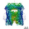 8764MC M: map data used to model this data C: citing same article ( |
|---|---|
| Similar structure data |
-Links
 PDBj
PDBj- Assembly
| Deposited unit | 
|
|---|---|
| 1 |
|
-Components
| #1: Protein | Mass: 65092.301 Da / Num. of mol.: 4 / Mutation: N138Q Source method: isolated from a genetically manipulated source Source: (gene. exp.) Gene: MCOLN3 / Plasmid: pFastBac / Cell line (production host): Sf9 / Production host:   Spodoptera frugiperda (fall armyworm) / References: UniProt: F6RG56 Spodoptera frugiperda (fall armyworm) / References: UniProt: F6RG56#2: Chemical | ChemComp-Y01 /   Mass: 486.726 Da / Num. of mol.: 12 / Source method: obtained synthetically / Formula: C31H50O4 Mass: 486.726 Da / Num. of mol.: 12 / Source method: obtained synthetically / Formula: C31H50O4#3: Chemical | ChemComp-NA /   Mass: 22.990 Da / Num. of mol.: 6 / Source method: obtained synthetically / Formula: Na Mass: 22.990 Da / Num. of mol.: 6 / Source method: obtained synthetically / Formula: Na#4: Chemical | ChemComp-3PE /   Mass: 748.065 Da / Num. of mol.: 4 / Source method: obtained synthetically / Formula: C41H82NO8P / Comment: phospholipid*YM Mass: 748.065 Da / Num. of mol.: 4 / Source method: obtained synthetically / Formula: C41H82NO8P / Comment: phospholipid*YM#5: Water | ChemComp-HOH / |  Mass: 18.015 Da / Num. of mol.: 4 / Source method: isolated from a natural source / Formula: H2O Mass: 18.015 Da / Num. of mol.: 4 / Source method: isolated from a natural source / Formula: H2OHas protein modification | N | |
|---|
-Experimental details
-Experiment
| Experiment | Method: ELECTRON MICROSCOPY |
|---|---|
| EM experiment | Aggregation state: PARTICLE / 3D reconstruction method: single particle reconstruction |
- Sample preparation
| Component | Name: Transient Receptor Potential Mucolipin 3 / Type: COMPLEX / Entity ID: #1 / Source: RECOMBINANT |
|---|---|
| Molecular weight | Value: 0.26 MDa / Experimental value: NO |
| Source (natural) | Organism: |
| Source (recombinant) | Organism: Spodoptera frugiperda (fall armyworm) / Strain: Sf9 / Plasmid: pFastBac |
| Buffer solution | pH: 7.4 |
| Specimen | Embedding applied: NO / Shadowing applied: NO / Staining applied: NO / Vitrification applied: YES |
| Vitrification | Cryogen name: ETHANE |
- Electron microscopy imaging
| Experimental equipment |  Model: Titan Krios / Image courtesy: FEI Company |
|---|---|
| Microscopy | Model: FEI TITAN KRIOS |
| Electron gun | Electron source:  FIELD EMISSION GUN / Accelerating voltage: 300 kV / Illumination mode: FLOOD BEAM FIELD EMISSION GUN / Accelerating voltage: 300 kV / Illumination mode: FLOOD BEAM |
| Electron lens | Mode: BRIGHT FIELD |
| Image recording | Average exposure time: 12 sec. / Electron dose: 63 e/Å2 / Detector mode: SUPER-RESOLUTION / Film or detector model: GATAN K2 SUMMIT (4k x 4k) |
- Processing
| Software | Name: PHENIX / Version: 1.11.1_2575: / Classification: refinement | ||||||||||||||||||||||||
|---|---|---|---|---|---|---|---|---|---|---|---|---|---|---|---|---|---|---|---|---|---|---|---|---|---|
| EM software | Name: PHENIX / Category: model refinement | ||||||||||||||||||||||||
| CTF correction | Type: PHASE FLIPPING AND AMPLITUDE CORRECTION | ||||||||||||||||||||||||
| Symmetry | Point symmetry: C4 (4 fold cyclic) | ||||||||||||||||||||||||
| 3D reconstruction | Resolution: 2.94 Å / Resolution method: FSC 0.143 CUT-OFF / Num. of particles: 104084 / Symmetry type: POINT | ||||||||||||||||||||||||
| Refine LS restraints |
|
