 Movie
Movie Controller

[English] 日本語
 Yorodumi
- PDB-5wpq: Cryo-EM structure of mammalian endolysosomal TRPML1 channel in na... -
+ Open data

- Basic information
| Entry | Database: PDB / ID: 5wpq | ||||||||||||||||||||||||||||||||||||||||||||||||||||||||||||||||||||||||
|---|---|---|---|---|---|---|---|---|---|---|---|---|---|---|---|---|---|---|---|---|---|---|---|---|---|---|---|---|---|---|---|---|---|---|---|---|---|---|---|---|---|---|---|---|---|---|---|---|---|---|---|---|---|---|---|---|---|---|---|---|---|---|---|---|---|---|---|---|---|---|---|---|---|
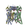
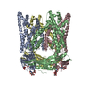
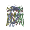
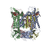
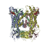
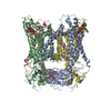
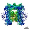
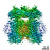
| Title | Cryo-EM structure of mammalian endolysosomal TRPML1 channel in nanodiscs in closed I conformation at 3.64 Angstrom resolution | ||||||||||||||||||||||||||||||||||||||||||||||||||||||||||||||||||||||||
 Components Components | Mucolipin-1 | ||||||||||||||||||||||||||||||||||||||||||||||||||||||||||||||||||||||||
 Keywords Keywords | MEMBRANE PROTEIN / Ion channel | ||||||||||||||||||||||||||||||||||||||||||||||||||||||||||||||||||||||||
| Function / homology |  Function and homology information Function and homology informationTransferrin endocytosis and recycling / positive regulation of lysosome organization / calcium ion export / intracellularly phosphatidylinositol-3,5-bisphosphate-gated monatomic cation channel activity / phagosome maturation / NAADP-sensitive calcium-release channel activity / iron ion transmembrane transporter activity / antibacterial innate immune response / iron ion transmembrane transport / cellular response to pH ...Transferrin endocytosis and recycling / positive regulation of lysosome organization / calcium ion export / intracellularly phosphatidylinositol-3,5-bisphosphate-gated monatomic cation channel activity / phagosome maturation / NAADP-sensitive calcium-release channel activity / iron ion transmembrane transporter activity / antibacterial innate immune response / iron ion transmembrane transport / cellular response to pH / TRP channels / monoatomic anion channel activity / ligand-gated calcium channel activity / negative regulation of protein localization to nucleus / endosomal transport / lysosome organization / sodium channel activity / intracellular vesicle / monoatomic cation transmembrane transport / autophagosome maturation / potassium channel activity / monoatomic cation channel activity / phagocytic cup / release of sequestered calcium ion into cytosol / negative regulation of innate immune response / cellular response to calcium ion / positive regulation of protein localization to nucleus / phagocytic vesicle membrane / calcium ion transmembrane transport / calcium channel activity / late endosome / late endosome membrane / protein homotetramerization / adaptive immune response / lysosome / signaling receptor complex / lysosomal membrane / lipid binding / Golgi apparatus / nucleoplasm / membrane / identical protein binding / plasma membrane Similarity search - Function | ||||||||||||||||||||||||||||||||||||||||||||||||||||||||||||||||||||||||
| Biological species |  | ||||||||||||||||||||||||||||||||||||||||||||||||||||||||||||||||||||||||
| Method | ELECTRON MICROSCOPY / single particle reconstruction / cryo EM / Resolution: 3.64 Å | ||||||||||||||||||||||||||||||||||||||||||||||||||||||||||||||||||||||||
 Authors Authors | Chen, Q. / She, J. / Guo, J. / Bai, X. / Jiang, Y. | ||||||||||||||||||||||||||||||||||||||||||||||||||||||||||||||||||||||||
| Funding support |  United States, 7items United States, 7items
| ||||||||||||||||||||||||||||||||||||||||||||||||||||||||||||||||||||||||
 Citation Citation | Journal: Nature / Year: 2017 Title: Structure of mammalian endolysosomal TRPML1 channel in nanodiscs. Authors: Qingfeng Chen / Ji She / Weizhong Zeng / Jiangtao Guo / Haoxing Xu / Xiao-Chen Bai / Youxing Jiang / Abstract: Transient receptor potential mucolipin 1 (TRPML1) is a cation channel located within endosomal and lysosomal membranes. Ubiquitously expressed in mammalian cells, its loss-of-function mutations are ...Transient receptor potential mucolipin 1 (TRPML1) is a cation channel located within endosomal and lysosomal membranes. Ubiquitously expressed in mammalian cells, its loss-of-function mutations are the direct cause of type IV mucolipidosis, an autosomal recessive lysosomal storage disease. Here we present the single-particle electron cryo-microscopy structure of the mouse TRPML1 channel embedded in nanodiscs. Combined with mutagenesis analysis, the TRPML1 structure reveals that phosphatidylinositol-3,5-bisphosphate (PtdIns(3,5)P) binds to the N terminus of the channel-distal from the pore-and the helix-turn-helix extension between segments S2 and S3 probably couples ligand binding to pore opening. The tightly packed selectivity filter contains multiple ion-binding sites, and the conserved acidic residues form the luminal Ca-blocking site that confers luminal pH and Ca modulation on channel conductance. A luminal linker domain forms a fenestrated canopy atop the channel, providing several luminal ion passages to the pore and creating a negative electrostatic trap, with a preference for divalent cations, at the luminal entrance. The structure also reveals two equally distributed S4-S5 linker conformations in the closed channel, suggesting an S4-S5 linker-mediated PtdInsP gating mechanism among TRPML channels. | ||||||||||||||||||||||||||||||||||||||||||||||||||||||||||||||||||||||||
| History |
|
- Structure visualization
| Movie |
Movie viewer |
|---|---|
| Structure viewer | Molecule: MolmilJmol/JSmol |
- Downloads & links
-Download
| PDBx/mmCIF format | 5wpq.cif.gz | 334.5 KB | Display | PDBx/mmCIF format |
|---|---|---|---|---|
| PDB format | pdb5wpq.ent.gz | 270.5 KB | Display | PDB format |
| PDBx/mmJSON format | 5wpq.json.gz | Tree view | PDBx/mmJSON format | |
| Others |  Other downloads Other downloads |
-Validation report
| Arichive directory | https://data.pdbj.org/pub/pdb/validation_reports/wp/5wpqftp://data.pdbj.org/pub/pdb/validation_reports/wp/5wpq | HTTPS FTP |
|---|
-Related structure data
| Related structure data | 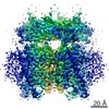 8881MC 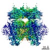 8882C 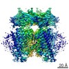 8883C 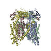 5wptC 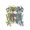 5wpvC  5wpo M: map data used to model this data C: citing same article ( |
|---|---|
| Similar structure data |

-Links
 PDBj
PDBj- Assembly
| Deposited unit | 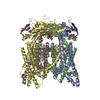
|
|---|---|
| 1 |
|
-Components
| #1: Protein | Mass: 66656.812 Da / Num. of mol.: 4 Source method: isolated from a genetically manipulated source Source: (gene. exp.)  Homo sapiens (human) / References: UniProt: Q99J21 Homo sapiens (human) / References: UniProt: Q99J21#2: Polysaccharide | 2-acetamido-2-deoxy-beta-D-glucopyranose-(1-4)-2-acetamido-2-deoxy-beta-D-glucopyranose Source method: isolated from a genetically manipulated source #3: Chemical | ChemComp-NA / |   Mass: 22.990 Da / Num. of mol.: 1 / Source method: obtained synthetically / Formula: Na Mass: 22.990 Da / Num. of mol.: 1 / Source method: obtained synthetically / Formula: NaHas protein modification | Y | |
|---|
-Experimental details
-Experiment
| Experiment | Method: ELECTRON MICROSCOPY |
|---|---|
| EM experiment | Aggregation state: PARTICLE / 3D reconstruction method: single particle reconstruction |
- Sample preparation
| Component | Name: Homotetramer of mouse TRPML1 / Type: ORGANELLE OR CELLULAR COMPONENT / Entity ID: #1 / Source: RECOMBINANT |
|---|---|
| Molecular weight | Value: 0.066 MDa / Experimental value: NO |
| Source (natural) | Organism: |
| Source (recombinant) | Organism: Homo sapiens (human) / Cell: HEK293F / Plasmid: pEZT |
| Buffer solution | pH: 8 Details: Solutions were made fresh from stock solutions and filtered. |
| Buffer component | Conc.: 150 mM / Name: sodium chloride / Formula: NaCl |
| Specimen | Conc.: 1.3 mg/ml / Embedding applied: NO / Shadowing applied: NO / Staining applied: NO / Vitrification applied: YES / Details: This sample was monodisperse |
| Specimen support | Grid material: COPPER / Grid mesh size: 200 divisions/in. / Grid type: Quantifoil R1.2/1.3 |
| Vitrification | Instrument: FEI VITROBOT MARK IV / Cryogen name: ETHANE / Humidity: 100 % / Chamber temperature: 277 K |
- Electron microscopy imaging
| Experimental equipment |  Model: Titan Krios / Image courtesy: FEI Company |
|---|---|
| Microscopy | Model: FEI TITAN KRIOS |
| Electron gun | Electron source:  FIELD EMISSION GUN / Accelerating voltage: 300 kV / Illumination mode: FLOOD BEAM FIELD EMISSION GUN / Accelerating voltage: 300 kV / Illumination mode: FLOOD BEAM |
| Electron lens | Mode: BRIGHT FIELD / Nominal magnification: 46730 X / Nominal defocus max: 3000 nm / Nominal defocus min: 1200 nm / Cs: 2.7 mm / C2 aperture diameter: 70 µm / Alignment procedure: COMA FREE |
| Specimen holder | Cryogen: NITROGEN / Specimen holder model: FEI TITAN KRIOS AUTOGRID HOLDER |
| Image recording | Average exposure time: 15 sec. / Electron dose: 50 e/Å2 / Detector mode: COUNTING / Film or detector model: GATAN K2 SUMMIT (4k x 4k) / Num. of grids imaged: 1 / Num. of real images: 3000 |
| EM imaging optics | Energyfilter name: GIFQuantum / Energyfilter upper: 10 eV / Energyfilter lower: -10 eV |
| Image scans | Movie frames/image: 30 |
- Processing
| Software | Name: PHENIX / Version: 1.11.1_2575: / Classification: refinement | ||||||||||||||||||||||||||||||||||||
|---|---|---|---|---|---|---|---|---|---|---|---|---|---|---|---|---|---|---|---|---|---|---|---|---|---|---|---|---|---|---|---|---|---|---|---|---|---|
| EM software |
| ||||||||||||||||||||||||||||||||||||
| Image processing | Details: 30 frames per movie stack were saved for motion correction. | ||||||||||||||||||||||||||||||||||||
| CTF correction | Details: The CTF correction was performed during the map refinement in RELION. Type: PHASE FLIPPING AND AMPLITUDE CORRECTION | ||||||||||||||||||||||||||||||||||||
| Particle selection | Num. of particles selected: 1433949 / Details: The particles were auto-picked in RELION. | ||||||||||||||||||||||||||||||||||||
| Symmetry | Point symmetry: C4 (4 fold cyclic) | ||||||||||||||||||||||||||||||||||||
| 3D reconstruction | Resolution: 3.64 Å / Resolution method: FSC 0.143 CUT-OFF / Num. of particles: 9000 / Algorithm: BACK PROJECTION / Num. of class averages: 2 / Symmetry type: 2D CRYSTAL | ||||||||||||||||||||||||||||||||||||
| Refine LS restraints |
|
