 Movie
Movie Controller


+ Open data

- Basic information
| Entry | Database: PDB / ID: 6e7y | ||||||||||||||||||||||||
|---|---|---|---|---|---|---|---|---|---|---|---|---|---|---|---|---|---|---|---|---|---|---|---|---|---|
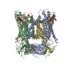
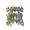
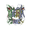
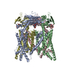
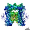
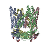
| Title | cryo-EM structure of human TRPML1 with PI45P2 | ||||||||||||||||||||||||
 Components Components | Mucolipin-1 | ||||||||||||||||||||||||
 Keywords Keywords | MEMBRANE PROTEIN / human TRPML1 | ||||||||||||||||||||||||
| Function / homology |  Function and homology information Function and homology informationpositive regulation of lysosome organization / calcium ion export / intracellularly phosphatidylinositol-3,5-bisphosphate-gated monatomic cation channel activity / phagosome maturation / NAADP-sensitive calcium-release channel activity / iron ion transmembrane transporter activity / iron ion transmembrane transport / cellular response to pH / Transferrin endocytosis and recycling / monoatomic anion channel activity ...positive regulation of lysosome organization / calcium ion export / intracellularly phosphatidylinositol-3,5-bisphosphate-gated monatomic cation channel activity / phagosome maturation / NAADP-sensitive calcium-release channel activity / iron ion transmembrane transporter activity / iron ion transmembrane transport / cellular response to pH / Transferrin endocytosis and recycling / monoatomic anion channel activity / intracellular zinc ion homeostasis / ligand-gated calcium channel activity / TRP channels / intracellular vesicle / sodium channel activity / autophagosome maturation / monoatomic cation transport / potassium channel activity / monoatomic cation channel activity / phagocytic cup / release of sequestered calcium ion into cytosol / cellular response to calcium ion / transferrin transport / phagocytic vesicle membrane / calcium channel activity / calcium ion transmembrane transport / late endosome / late endosome membrane / protein homotetramerization / adaptive immune response / lysosome / signaling receptor complex / endosome membrane / lysosomal membrane / lipid binding / Golgi apparatus / nucleoplasm / membrane / identical protein binding / plasma membrane Similarity search - Function | ||||||||||||||||||||||||
| Biological species |  Homo sapiens (human) Homo sapiens (human) | ||||||||||||||||||||||||
| Method | ELECTRON MICROSCOPY / single particle reconstruction / cryo EM / Resolution: 3.57 Å | ||||||||||||||||||||||||
 Authors Authors | Schmiege, P. / Li, X. | ||||||||||||||||||||||||
 Citation Citation | Journal: Nat Commun / Year: 2018 Title: Structural basis for PtdInsP-mediated human TRPML1 regulation. Authors: Michael Fine / Philip Schmiege / Xiaochun Li /  Abstract: Transient receptor potential mucolipin 1 (TRPML1), a lysosomal channel, maintains the low pH and calcium levels for lysosomal function. Several small molecules modulate TRPML1 activity. ML-SA1, a ...Transient receptor potential mucolipin 1 (TRPML1), a lysosomal channel, maintains the low pH and calcium levels for lysosomal function. Several small molecules modulate TRPML1 activity. ML-SA1, a synthetic agonist, binds to the pore region and phosphatidylinositol-3,5-bisphosphate (PtdIns(3,5)P), a natural lipid, stimulates channel activity to a lesser extent than ML-SA1; moreover, PtdIns(4,5)P, another natural lipid, prevents TRPML1-mediated calcium release. Notably, PtdIns(3,5)P and ML-SA1 cooperate further increasing calcium efflux. Here we report the structures of human TRPML1 at pH 5.0 with PtdIns(3,5)P, PtdIns(4,5)P, or ML-SA1 and PtdIns(3,5)P, revealing a unique lipid-binding site. PtdIns(3,5)P and PtdIns(4,5)P bind to the extended helices of S1, S2, and S3. The phosphate group of PtdIns(3,5)P induces Y355 to form a π-cation interaction with R403, moving the S4-S5 linker, thus allosterically activating the channel. Our structures and electrophysiological characterizations reveal an allosteric site and provide molecular insight into how lipids regulate TRP channels. | ||||||||||||||||||||||||
| History |
|
- Structure visualization
| Movie |
Movie viewer |
|---|---|
| Structure viewer | Molecule: MolmilJmol/JSmol |
- Downloads & links
-Download
| PDBx/mmCIF format | 6e7y.cif.gz | 340.5 KB | Display | PDBx/mmCIF format |
|---|---|---|---|---|
| PDB format | pdb6e7y.ent.gz | 280.6 KB | Display | PDB format |
| PDBx/mmJSON format | 6e7y.json.gz | Tree view | PDBx/mmJSON format | |
| Others |  Other downloads Other downloads |
-Validation report
| Arichive directory | https://data.pdbj.org/pub/pdb/validation_reports/e7/6e7yftp://data.pdbj.org/pub/pdb/validation_reports/e7/6e7y | HTTPS FTP |
|---|
-Related structure data
| Related structure data | 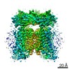 9001MC 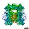 9000C 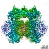 9002C 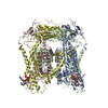 6e7pC 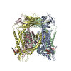 6e7zC M: map data used to model this data C: citing same article ( |
|---|---|
| Similar structure data |

-Links
 PDBj
PDBj
- Assembly
| Deposited unit | 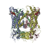
|
|---|---|
| 1 |
|
-Components
| #1: Protein | Mass: 65084.996 Da / Num. of mol.: 4 Source method: isolated from a genetically manipulated source Source: (gene. exp.) Homo sapiens (human) / Gene: MCOLN1, ML4, MSTP080 / Production host: Homo sapiens (human) / References: UniProt: Q9GZU1#2: Chemical | ChemComp-PIO / [(   Mass: 746.566 Da / Num. of mol.: 4 / Source method: obtained synthetically / Formula: C25H49O19P3 Mass: 746.566 Da / Num. of mol.: 4 / Source method: obtained synthetically / Formula: C25H49O19P3Has protein modification | Y | |
|---|
-Experimental details
-Experiment
| Experiment | Method: ELECTRON MICROSCOPY |
|---|---|
| EM experiment | Aggregation state: PARTICLE / 3D reconstruction method: single particle reconstruction |
- Sample preparation
| Component | Name: TRPML1 / Type: COMPLEX / Entity ID: #1 / Source: RECOMBINANT |
|---|---|
| Molecular weight | Experimental value: NO |
| Source (natural) | Organism: Homo sapiens (human) |
| Source (recombinant) | Organism: Homo sapiens (human) |
| Buffer solution | pH: 7 |
| Specimen | Conc.: 7 mg/ml / Embedding applied: NO / Shadowing applied: NO / Staining applied: NO / Vitrification applied: YES |
| Specimen support | Details: unspecified |
| Vitrification | Cryogen name: NITROGEN |
- Electron microscopy imaging
| Experimental equipment |  Model: Titan Krios / Image courtesy: FEI Company |
|---|---|
| Microscopy | Model: FEI TITAN KRIOS |
| Electron gun | Electron source:  FIELD EMISSION GUN / Accelerating voltage: 300 kV / Illumination mode: FLOOD BEAM FIELD EMISSION GUN / Accelerating voltage: 300 kV / Illumination mode: FLOOD BEAM |
| Electron lens | Mode: BRIGHT FIELD |
| Image recording | Electron dose: 1.6 e/Å2 / Film or detector model: GATAN K2 SUMMIT (4k x 4k) |
- Processing
| Software | Name: PHENIX / Version: 1.13_2998: / Classification: refinement | ||||||||||||||||||||||||
|---|---|---|---|---|---|---|---|---|---|---|---|---|---|---|---|---|---|---|---|---|---|---|---|---|---|
| EM software |
| ||||||||||||||||||||||||
| CTF correction | Type: PHASE FLIPPING AND AMPLITUDE CORRECTION | ||||||||||||||||||||||||
| 3D reconstruction | Resolution: 3.57 Å / Resolution method: FSC 0.143 CUT-OFF / Num. of particles: 69587 / Symmetry type: POINT | ||||||||||||||||||||||||
| Refinement | Highest resolution: 3.57 Å | ||||||||||||||||||||||||
| Refine LS restraints |
|
