 Movie
Movie Controller


+ Open data

- Basic information
| Entry | Database: PDB / ID: 5vvj | ||||||
|---|---|---|---|---|---|---|---|
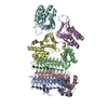
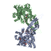
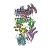
| Title | Cas1-Cas2 bound to half-site intermediate | ||||||
 Components Components |
| ||||||
 Keywords Keywords | HYDROLASE/DNA / Complex / DNA / HYDROLASE-DNA complex | ||||||
| Function / homology |  Function and homology information Function and homology informationCRISPR-cas system / crossover junction DNA endonuclease activity / 5'-flap endonuclease activity / maintenance of CRISPR repeat elements / endonuclease activity / defense response to virus / Hydrolases; Acting on ester bonds / hydrolase activity / DNA repair / DNA damage response ...CRISPR-cas system / crossover junction DNA endonuclease activity / 5'-flap endonuclease activity / maintenance of CRISPR repeat elements / endonuclease activity / defense response to virus / Hydrolases; Acting on ester bonds / hydrolase activity / DNA repair / DNA damage response / protein homodimerization activity / DNA binding / metal ion binding / identical protein binding / cytoplasm Similarity search - Function | ||||||
| Biological species |  synthetic construct (others) | ||||||
| Method |  X-RAY DIFFRACTION / SYNCHROTRON / MOLECULAR REPLACEMENT / molecular replacement / Resolution: 3.89 Å X-RAY DIFFRACTION / SYNCHROTRON / MOLECULAR REPLACEMENT / molecular replacement / Resolution: 3.89 Å | ||||||
 Authors Authors | Wright, A.V. / Knott, G.J. / Doxzen, K.W. / Doudna, J.A. | ||||||
| Funding support |  United States, 1items United States, 1items
| ||||||
 Citation Citation | Journal: Science / Year: 2017 Title: Structures of the CRISPR genome integration complex. Authors: Addison V Wright / Jun-Jie Liu / Gavin J Knott / Kevin W Doxzen / Eva Nogales / Jennifer A Doudna / Abstract: CRISPR-Cas systems depend on the Cas1-Cas2 integrase to capture and integrate short foreign DNA fragments into the CRISPR locus, enabling adaptation to new viruses. We present crystal structures of ...CRISPR-Cas systems depend on the Cas1-Cas2 integrase to capture and integrate short foreign DNA fragments into the CRISPR locus, enabling adaptation to new viruses. We present crystal structures of Cas1-Cas2 bound to both donor and target DNA in intermediate and product integration complexes, as well as a cryo-electron microscopy structure of the full CRISPR locus integration complex, including the accessory protein IHF (integration host factor). The structures show unexpectedly that indirect sequence recognition dictates integration site selection by favoring deformation of the repeat and the flanking sequences. IHF binding bends the DNA sharply, bringing an upstream recognition motif into contact with Cas1 to increase both the specificity and efficiency of integration. These results explain how the Cas1-Cas2 CRISPR integrase recognizes a sequence-dependent DNA structure to ensure site-selective CRISPR array expansion during the initial step of bacterial adaptive immunity. | ||||||
| History |
|
- Structure visualization
| Structure viewer | Molecule: MolmilJmol/JSmol |
|---|
- Downloads & links
-Download
| PDBx/mmCIF format | 5vvj.cif.gz | 615.3 KB | Display | PDBx/mmCIF format |
|---|---|---|---|---|
| PDB format | pdb5vvj.ent.gz | 506.8 KB | Display | PDB format |
| PDBx/mmJSON format | 5vvj.json.gz | Tree view | PDBx/mmJSON format | |
| Others |  Other downloads Other downloads |
-Validation report
| Arichive directory | https://data.pdbj.org/pub/pdb/validation_reports/vv/5vvjftp://data.pdbj.org/pub/pdb/validation_reports/vv/5vvj | HTTPS FTP |
|---|
-Related structure data
| Related structure data | 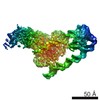 8827C 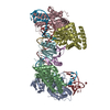 5vvkC 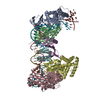 5vvlC 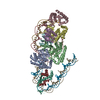 5wfeC  5ds5S S: Starting model for refinement C: citing same article ( |
|---|---|
| Similar structure data |


-Links
 PDBj
PDBj


- Assembly
| Deposited unit | 
| ||||||||
|---|---|---|---|---|---|---|---|---|---|
| 1 |
| ||||||||
| Unit cell |
|
-Components
| #1: Protein | Mass: 33235.418 Da / Num. of mol.: 4 Source method: isolated from a genetically manipulated source Source: (gene. exp.) Strain: K12 / Gene: ygbT, cas1, b2755, JW2725 / Variant: MG1655 / Production host: References: UniProt: Q46896, Hydrolases; Acting on ester bonds #2: Protein | Mass: 10584.264 Da / Num. of mol.: 2 Source method: isolated from a genetically manipulated source Source: (gene. exp.) Strain: K12 / Gene: ygbF, cas2, b2754, JW5438 / Variant: MG1655 / Production host: References: UniProt: P45956, Hydrolases; Acting on ester bonds #3: DNA chain | | Mass: 8680.646 Da / Num. of mol.: 1 / Source method: obtained synthetically / Source: (synth.) synthetic construct (others) #4: DNA chain | | Mass: 34538.016 Da / Num. of mol.: 1 / Source method: obtained synthetically / Source: (synth.) synthetic construct (others) |
|---|
-Experimental details
-Experiment
| Experiment | Method: X-RAY DIFFRACTION / Number of used crystals: 1 |
|---|
- Sample preparation
| Crystal | Density Matthews: 3.43 Å3/Da / Density % sol: 64.13 % |
|---|---|
| Crystal grow | Temperature: 281 K / Method: vapor diffusion, hanging drop / pH: 6.5 Details: 100 mM MES, pH 6.5, 10% (w/v) poly(ethylene glycol) (PEG) monomethyl ether (MME) 5000, 12% (v/v) propanol |
-Data collection
| Diffraction | Mean temperature: 80 K | |||||||||||||||||||||
|---|---|---|---|---|---|---|---|---|---|---|---|---|---|---|---|---|---|---|---|---|---|---|
| Diffraction source | Source: SYNCHROTRON / Site: ALS / Beamline: 8.3.1 / Wavelength: 1.1158 Å | |||||||||||||||||||||
| Detector | Type: DECTRIS PILATUS3 S 6M / Detector: PIXEL / Date: Apr 20, 2017 | |||||||||||||||||||||
| Radiation | Protocol: SINGLE WAVELENGTH / Monochromatic (M) / Laue (L): M / Scattering type: x-ray | |||||||||||||||||||||
| Radiation wavelength | Wavelength: 1.1158 Å / Relative weight: 1 | |||||||||||||||||||||
| Reflection | Resolution: 3.28→196.92 Å / Num. obs: 40983 / % possible obs: 96.1 % / Redundancy: 12.3 % / Biso Wilson estimate: 126.51 Å2 / CC1/2: 0.998 / Rmerge(I) obs: 0.668 / Rpim(I) all: 0.195 / Rrim(I) all: 0.697 / Net I/σ(I): 3.3 | |||||||||||||||||||||
| Reflection shell | Diffraction-ID: 1
|
-Phasing
| Phasing | Method: molecular replacement |
|---|
- Processing
| Software |
| ||||||||||||||||||||||||||||||||||||||||||||||||||||||||||||
|---|---|---|---|---|---|---|---|---|---|---|---|---|---|---|---|---|---|---|---|---|---|---|---|---|---|---|---|---|---|---|---|---|---|---|---|---|---|---|---|---|---|---|---|---|---|---|---|---|---|---|---|---|---|---|---|---|---|---|---|---|---|
| Refinement | Method to determine structure: MOLECULAR REPLACEMENT Starting model: 5DS5 Resolution: 3.89→134.096 Å / SU ML: 0.7 / Cross valid method: FREE R-VALUE / σ(F): 1.34 / Phase error: 38.09
| ||||||||||||||||||||||||||||||||||||||||||||||||||||||||||||
| Solvent computation | Shrinkage radii: 0.9 Å / VDW probe radii: 1.11 Å | ||||||||||||||||||||||||||||||||||||||||||||||||||||||||||||
| Displacement parameters | Biso max: 549.16 Å2 / Biso mean: 158.4474 Å2 / Biso min: 52.96 Å2 | ||||||||||||||||||||||||||||||||||||||||||||||||||||||||||||
| Refinement step | Cycle: final / Resolution: 3.89→134.096 Å
| ||||||||||||||||||||||||||||||||||||||||||||||||||||||||||||
| Refine LS restraints |
| ||||||||||||||||||||||||||||||||||||||||||||||||||||||||||||
| LS refinement shell | Refine-ID: X-RAY DIFFRACTION / Rfactor Rfree error: 0 / Total num. of bins used: 9 / % reflection obs: 100 %
|
