 Movie
Movie Controller

[English] 日本語
 Yorodumi
- PDB-3weu: Crystal structure of the L-Lys epsilon-oxidase from Marinomonas m... -
+ Open data

- Basic information
| Entry | Database: PDB / ID: 3weu | ||||||
|---|---|---|---|---|---|---|---|
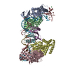
| Title | Crystal structure of the L-Lys epsilon-oxidase from Marinomonas mediterranea | ||||||
 Components Components | L-lysine 6-oxidase | ||||||
 Keywords Keywords | OXIDOREDUCTASE / amino acid oxidase / arm / beta barrel / three beta sheets / amine oxidase / L-Lys binding | ||||||
| Function / homology |  Function and homology information Function and homology informationL-lysine 6-oxidase / L-lysine 6-oxidase activity / negative regulation of single-species biofilm formation / quinone binding / killing of cells of another organism / defense response to bacterium / extracellular region Similarity search - Function | ||||||
| Biological species |  Marinomonas mediterranea (bacteria) Marinomonas mediterranea (bacteria) | ||||||
| Method |  X-RAY DIFFRACTION / SYNCHROTRON / SAD / Resolution: 1.93 Å X-RAY DIFFRACTION / SYNCHROTRON / SAD / Resolution: 1.93 Å | ||||||
 Authors Authors | Okazaki, S. / Nakano, S. / Matsui, D. / Akaji, S. / Inagaki, K. / Asano, Y. | ||||||
 Citation Citation | Journal: J.Biochem. / Year: 2013 Title: X-Ray crystallographic evidence for the presence of the cysteine tryptophylquinone cofactor in L-lysine {varepsilon}-oxidase from Marinomonas mediterranea Authors: Okazaki, S. / Nakano, S. / Matsui, D. / Akaji, S. / Inagaki, K. / Asano, Y. | ||||||
| History |
|
- Structure visualization
| Structure viewer | Molecule: MolmilJmol/JSmol |
|---|
- Downloads & links
-Download
| PDBx/mmCIF format | 3weu.cif.gz | 309.5 KB | Display | PDBx/mmCIF format |
|---|---|---|---|---|
| PDB format | pdb3weu.ent.gz | 250 KB | Display | PDB format |
| PDBx/mmJSON format | 3weu.json.gz | Tree view | PDBx/mmJSON format | |
| Others |  Other downloads Other downloads |
-Validation report
| Arichive directory | https://data.pdbj.org/pub/pdb/validation_reports/we/3weuftp://data.pdbj.org/pub/pdb/validation_reports/we/3weu | HTTPS FTP |
|---|
-Related structure data


-Links
 PDBj
PDBj- Assembly
| Deposited unit | 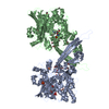
| |||||||||
|---|---|---|---|---|---|---|---|---|---|---|
| 1 |
| |||||||||
| 2 | 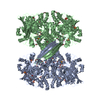
| |||||||||
| Unit cell |
| |||||||||
| Components on special symmetry positions |
|
-Components
| #1: Protein | Mass: 81010.219 Da / Num. of mol.: 2 Source method: isolated from a genetically manipulated source Source: (gene. exp.) Marinomonas mediterranea (bacteria) / Strain: NBRC 103028 / Gene: lodA / Production host: Marinomonas mediterranea (bacteria) / Strain (production host): NBRC 103028 / References: UniProt: F2JXJ3, L-lysine 6-oxidase#2: Chemical | ChemComp-SO4 /   Mass: 96.063 Da / Num. of mol.: 6 / Source method: obtained synthetically / Formula: SO4 Mass: 96.063 Da / Num. of mol.: 6 / Source method: obtained synthetically / Formula: SO4#3: Chemical |   Mass: 88.105 Da / Num. of mol.: 2 / Source method: obtained synthetically / Formula: C4H8O2 Mass: 88.105 Da / Num. of mol.: 2 / Source method: obtained synthetically / Formula: C4H8O2#4: Chemical | ChemComp-EDO /   Mass: 62.068 Da / Num. of mol.: 18 / Source method: obtained synthetically / Formula: C2H6O2 Mass: 62.068 Da / Num. of mol.: 18 / Source method: obtained synthetically / Formula: C2H6O2#5: Water | ChemComp-HOH / |  Mass: 18.015 Da / Num. of mol.: 1128 / Source method: isolated from a natural source / Formula: H2O Mass: 18.015 Da / Num. of mol.: 1128 / Source method: isolated from a natural source / Formula: H2OHas protein modification | Y | |
|---|
-Experimental details
-Experiment
| Experiment | Method: X-RAY DIFFRACTION / Number of used crystals: 1 |
|---|
- Sample preparation
| Crystal | Density Matthews: 2.62 Å3/Da / Density % sol: 53.12 % |
|---|---|
| Crystal grow | Temperature: 293 K / Method: vapor diffusion, sitting drop / pH: 6.5 Details: 1.6M ammonium sulfate, 10%(v/v) 1,4-dioxane, 0.1M MES/NaOH, 0.2M NaI, pH 6.5, VAPOR DIFFUSION, SITTING DROP, temperature 293K |
-Data collection
| Diffraction | Mean temperature: 100 K |
|---|---|
| Diffraction source | Source: SYNCHROTRON / Site: Photon Factory  / Beamline: BL-1A / Wavelength: 1.1 Å / Beamline: BL-1A / Wavelength: 1.1 Å |
| Detector | Type: DECTRIS PILATUS 2M-F / Detector: PIXEL / Date: Feb 20, 2013 |
| Radiation | Monochromator: Cryo-cooled channel-cut Si(111) / Protocol: SINGLE WAVELENGTH / Monochromatic (M) / Laue (L): M / Scattering type: x-ray |
| Radiation wavelength | Wavelength: 1.1 Å / Relative weight: 1 |
| Reflection | Resolution: 1.93→50 Å / Num. obs: 124499 / % possible obs: 99.4 % / Observed criterion σ(F): 1 / Observed criterion σ(I): 1 / Redundancy: 13.1 % / Rmerge(I) obs: 0.065 / Net I/σ(I): 13.1 |
| Reflection shell | Resolution: 1.94→1.97 Å / Redundancy: 9.6 % / Rmerge(I) obs: 0.24 / Mean I/σ(I) obs: 6.8 / Num. unique all: 21819 / % possible all: 92.5 |
- Processing
| Software |
| ||||||||||||||||||||||||||||||||||||||||||||||||||||||||||||||||||||||||||||||||||||||||||||||||||||||||||||||||||||||||||||||||||||||||||||||||||||||||||||||||||||||||||||||||||||||
|---|---|---|---|---|---|---|---|---|---|---|---|---|---|---|---|---|---|---|---|---|---|---|---|---|---|---|---|---|---|---|---|---|---|---|---|---|---|---|---|---|---|---|---|---|---|---|---|---|---|---|---|---|---|---|---|---|---|---|---|---|---|---|---|---|---|---|---|---|---|---|---|---|---|---|---|---|---|---|---|---|---|---|---|---|---|---|---|---|---|---|---|---|---|---|---|---|---|---|---|---|---|---|---|---|---|---|---|---|---|---|---|---|---|---|---|---|---|---|---|---|---|---|---|---|---|---|---|---|---|---|---|---|---|---|---|---|---|---|---|---|---|---|---|---|---|---|---|---|---|---|---|---|---|---|---|---|---|---|---|---|---|---|---|---|---|---|---|---|---|---|---|---|---|---|---|---|---|---|---|---|---|---|---|
| Refinement | Method to determine structure: SAD / Resolution: 1.93→47.54 Å / Cor.coef. Fo:Fc: 0.967 / Cor.coef. Fo:Fc free: 0.951 / SU B: 2.329 / SU ML: 0.069 / Cross valid method: THROUGHOUT / ESU R: 0.114 / ESU R Free: 0.11 / Stereochemistry target values: MAXIMUM LIKELIHOOD / Details: HYDROGENS HAVE BEEN ADDED IN THE RIDING POSITIONS
| ||||||||||||||||||||||||||||||||||||||||||||||||||||||||||||||||||||||||||||||||||||||||||||||||||||||||||||||||||||||||||||||||||||||||||||||||||||||||||||||||||||||||||||||||||||||
| Solvent computation | Ion probe radii: 0.8 Å / Shrinkage radii: 0.8 Å / VDW probe radii: 1.2 Å / Solvent model: MASK | ||||||||||||||||||||||||||||||||||||||||||||||||||||||||||||||||||||||||||||||||||||||||||||||||||||||||||||||||||||||||||||||||||||||||||||||||||||||||||||||||||||||||||||||||||||||
| Displacement parameters | Biso mean: 21.737 Å2
| ||||||||||||||||||||||||||||||||||||||||||||||||||||||||||||||||||||||||||||||||||||||||||||||||||||||||||||||||||||||||||||||||||||||||||||||||||||||||||||||||||||||||||||||||||||||
| Refinement step | Cycle: LAST / Resolution: 1.93→47.54 Å
| ||||||||||||||||||||||||||||||||||||||||||||||||||||||||||||||||||||||||||||||||||||||||||||||||||||||||||||||||||||||||||||||||||||||||||||||||||||||||||||||||||||||||||||||||||||||
| Refine LS restraints |
|