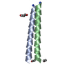
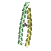
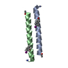
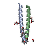
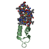
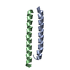
Entry Database : PDB / ID : 5vr3Title Crystal structure of the BRS domain of BRAF BRAF Keywords / Function / homology Function Domain/homology Component
/ / / / / / / / / / / / / / / / / / / / / / / / / / / / / / / / / / / / / / / / / / / / / / / / / / / / / / / / / / / / / / / / / / / / / / / / / / / / / / / / / / / / / / / / / / / / / / / / / / / / / / / / / / / / / / / / / / / / / / / / / / / / / / / Biological species Homo sapiens (human)Method / / / Resolution : 2.102 Å Authors Thevakumaran, N. / Maisonneuve, P. / Kurinov, I. / Lavoie, H. / Marullo, S.A. / Sahmi, M. / Jin, T. / Therrien, M. / Sicheri, F. Journal : Nature / Year : 2018Title : MEK drives BRAF activation through allosteric control of KSR proteins.Authors : Lavoie, H. / Sahmi, M. / Maisonneuve, P. / Marullo, S.A. / Thevakumaran, N. / Jin, T. / Kurinov, I. / Sicheri, F. / Therrien, M. History Deposition May 10, 2017 Deposition site / Processing site Revision 1.0 Feb 14, 2018 Provider / Type Revision 1.1 Feb 28, 2018 Group / Category / citation_authorItem / _citation.title / _citation_author.nameRevision 1.2 Mar 7, 2018 Group / Category Item / _citation.page_first / _citation.page_lastRevision 1.3 Mar 13, 2024 Group / Database references / Category / chem_comp_bond / database_2Item / _database_2.pdbx_database_accession
Show all Show less
 Movie
Movie Controller


 Open data
 Basic information
 Components
 Keywords
 Function and homology information
 Homo sapiens (human)
 X-RAY DIFFRACTION /
  Authors
 Citation
 Structure visualization
 Downloads & links
 Other downloads
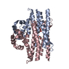


 PDBj
PDBj


 Assembly


 Mass: 96.063 Da / Num. of mol.: 1 / Source method: obtained synthetically / Formula: SO4
 Mass: 18.015 Da / Num. of mol.: 13 / Source method: isolated from a natural source / Formula: H2O
 Sample preparation
 / Beamline: 24-ID-C / Wavelength: 0.9792 Å
 Processing