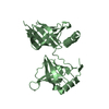
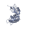
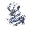
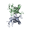
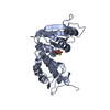
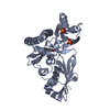
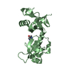
[English] 日本語
Yorodumi
- PDB-5vqh: Crystal structure of the extended Tudor domain from BmPAPI in com... -

+
Open data


ID or keywords:

Loading...

-
Basic information

Entry
Database: PDB / ID: 5vqh
TitleCrystal structure of the extended Tudor domain from BmPAPI in complex with sDMA
ComponentsTudor and KH domain-containing protein homolog
KeywordsRNA BINDING PROTEIN / BmPAPI / Tudor / sDMA
Function / homology
Function and homology information


piRNA processing / spermatogenesis / mitochondrial outer membrane / cell differentiation / RNA binding
Similarity search - Function
: / Tudor domain / Tudor domain profile. / KH domain / K Homology domain, type 1 / Tudor domain / Tudor domain / Type-1 KH domain profile. / OB fold (Dihydrolipoamide Acetyltransferase, E2P) - #90 / SH3 type barrels. - #140 ...: / Tudor domain / Tudor domain profile. / KH domain / K Homology domain, type 1 / Tudor domain / Tudor domain / Type-1 KH domain profile. / OB fold (Dihydrolipoamide Acetyltransferase, E2P) - #90 / SH3 type barrels. - #140 / SNase-like, OB-fold superfamily / K Homology domain, type 1 superfamily / K Homology domain / K homology RNA-binding domain / SH3 type barrels. / OB fold (Dihydrolipoamide Acetyltransferase, E2P) / Roll / Beta Barrel / Mainly Beta
Similarity search - Domain/homology
N3, N4-DIMETHYLARGININE / Tudor and KH domain-containing protein homolog
Similarity search - Component
Biological speciesBombyx mori (domestic silkworm)
MethodX-RAY DIFFRACTION / MOLECULAR REPLACEMENT / Resolution: 2.4 Å
AuthorsHubbard, P.A. / Pan, X. / Ohtaki, A. / McNally, R. / Honda, S. / Kirino, Y. / Murali, R.
Funding support United States, 1items
OrganizationGrant numberCountry
National Institutes of Health/National Institute of General Medical Sciences (NIH/NIGMS)GM106047 United States
CitationJournal: To Be Published
Title: Structural studies of the Tudor domain from the Bombyx homolog of Drosophila PAPI: Implication to piRNA biogenesis
Authors: Hubbard, P.A. / Pan, X. / Ohtaki, A. / McNally, R. / Honda, S. / Kirino, Y. / Murali, R.
History
DepositionMay 8, 2017Deposition site: RCSB / Processing site: RCSB
Revision 1.0Jun 14, 2017Provider: repository / Type: Initial release
Revision 1.1Sep 13, 2017Group: Author supporting evidence / Category: pdbx_audit_support / Item: _pdbx_audit_support.funding_organization
Revision 1.2Jan 1, 2020Group: Author supporting evidence / Derived calculations / Category: pdbx_audit_support / struct_conn
Item: _pdbx_audit_support.funding_organization / _struct_conn.pdbx_leaving_atom_flag
Revision 1.3Mar 10, 2021Group: Derived calculations / Category: struct_conn
Revision 1.4Oct 4, 2023Group: Data collection / Database references / Refinement description
Category: chem_comp_atom / chem_comp_bond ...chem_comp_atom / chem_comp_bond / database_2 / pdbx_initial_refinement_model
Item: _database_2.pdbx_DOI / _database_2.pdbx_database_accession

-
Structure visualization

Structure viewerMolecule:
MolmilJmol/JSmol

Downloads & links

-
Assembly

Deposited unit
A: Tudor and KH domain-containing protein homolog
B: Tudor and KH domain-containing protein homolog
hetero molecules


Theoretical massNumber of molelcules
Total (without water)54,3114
Polymers53,9062
Non-polymers4052
Water2,756153
1
A: Tudor and KH domain-containing protein homolog
hetero molecules


Theoretical massNumber of molelcules
Total (without water)27,1552
Polymers26,9531
Non-polymers2021
Water181
TypeNameSymmetry operationNumber
identity operation1_555x,y,z1
2
B: Tudor and KH domain-containing protein homolog
hetero molecules


Theoretical massNumber of molelcules
Total (without water)27,1552
Polymers26,9531
Non-polymers2021
Water181
TypeNameSymmetry operationNumber
identity operation1_555x,y,z1
Unit cell
Length a, b, c (Å)66.030, 66.030, 224.480
Angle α, β, γ (deg.)90.00, 90.00, 90.00
Int Tables number96
Space group name H-MP43212
Components on special symmetry positions
IDModelComponents
11A-614-

HOH

-
Components

#1: Protein Tudor and KH domain-containing protein homolog / Partner of PIWIs protein / BmPAPI


Mass: 26953.027 Da / Num. of mol.: 2 / Fragment: UNP residues 245-462
Source method: isolated from a genetically manipulated source
Source: (gene. exp.) Bombyx mori (domestic silkworm) / Gene: PAPI / Production host: Escherichia coli (E. coli) / References: UniProt: H9JD76
#2: Chemical ChemComp-2MR / N3, N4-DIMETHYLARGININE


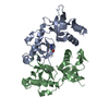
Type: L-peptide linking / Mass: 202.254 Da / Num. of mol.: 2
Source method: isolated from a genetically manipulated source
Formula: C8H18N4O2
#3: Water ChemComp-HOH / water / Water


Mass: 18.015 Da / Num. of mol.: 153 / Source method: isolated from a natural source / Formula: H2O

-
Experimental details

-
Experiment

ExperimentMethod: X-RAY DIFFRACTION / Number of used crystals: 1

-
Sample preparation

CrystalDensity Matthews: 2.25 Å3/Da / Density % sol: 45.4 %
Crystal growTemperature: 281 K / Method: vapor diffusion, hanging drop
Details: 0.95 M ammonium sulfate, 50 mM sodium acetate, pH 5.5 and 5% glucose.

-
Data collection

DiffractionMean temperature: 100 K
Diffraction sourceSource: ROTATING ANODE / Type: RIGAKU MICROMAX-007 HF / Wavelength: 1.5418 Å
DetectorType: RIGAKU / Detector: IMAGE PLATE / Date: Jun 25, 2015
RadiationProtocol: SINGLE WAVELENGTH / Monochromatic (M) / Laue (L): M / Scattering type: x-ray
Radiation wavelengthWavelength: 1.5418 Å / Relative weight: 1
ReflectionResolution: 2.4→39.61 Å / Num. obs: 18600 / % possible obs: 92.8 % / Redundancy: 3.7 % / Rmerge(I) obs: 0.077 / Net I/σ(I): 8.7
Reflection shellResolution: 2.4→2.51 Å / Redundancy: 3 % / Rmerge(I) obs: 0.535 / Mean I/σ(I) obs: 1.6 / Num. unique all: 6867 / % possible all: 95

-
Processing

Software
NameVersionClassification
REFMAC5.8.0073refinement
iMOSFLMdata reduction
XDSdata scaling
PHASERphasing
RefinementMethod to determine structure: MOLECULAR REPLACEMENT
Starting model: 5VQG
Resolution: 2.4→39.61 Å / Cor.coef. Fo:Fc: 0.949 / Cor.coef. Fo:Fc free: 0.909 / SU B: 10.461 / SU ML: 0.23 / Cross valid method: THROUGHOUT / ESU R: 0.448 / ESU R Free: 0.289 / Details: HYDROGENS HAVE BEEN ADDED IN THE RIDING POSITIONS
RfactorNum. reflection% reflectionSelection details
Rfree0.262 878 4.7 %RANDOM
Rwork0.195 ---
obs0.198 17693 91.1 %-
Solvent computationIon probe radii: 0.8 Å / Shrinkage radii: 0.8 Å / VDW probe radii: 1.2 Å
Displacement parametersBiso mean: 45.17 Å2
Baniso -1Baniso -2Baniso -3
1--0.36 Å20 Å20 Å2
2---0.36 Å20 Å2
3---0.72 Å2
Refinement stepCycle: LAST / Resolution: 2.4→39.61 Å
ProteinNucleic acidLigandSolventTotal
Num. atoms3213 0 28 153 3394
Refine LS restraints
Refine-IDTypeDev idealDev ideal targetNumber
X-RAY DIFFRACTIONr_bond_refined_d0.0120.0193330
X-RAY DIFFRACTIONr_bond_other_d0.0010.023058
X-RAY DIFFRACTIONr_angle_refined_deg1.4821.9324530
X-RAY DIFFRACTIONr_angle_other_deg0.77436971
X-RAY DIFFRACTIONr_dihedral_angle_1_deg6.9495407
X-RAY DIFFRACTIONr_dihedral_angle_2_deg32.69723.212165
X-RAY DIFFRACTIONr_dihedral_angle_3_deg15.05215498
X-RAY DIFFRACTIONr_dihedral_angle_4_deg20.021526
X-RAY DIFFRACTIONr_chiral_restr0.0790.2496
X-RAY DIFFRACTIONr_gen_planes_refined0.0050.023818
X-RAY DIFFRACTIONr_gen_planes_other0.0010.02826
X-RAY DIFFRACTIONr_nbd_refined
X-RAY DIFFRACTIONr_nbd_other
X-RAY DIFFRACTIONr_nbtor_refined
X-RAY DIFFRACTIONr_nbtor_other
X-RAY DIFFRACTIONr_xyhbond_nbd_refined
X-RAY DIFFRACTIONr_xyhbond_nbd_other
X-RAY DIFFRACTIONr_metal_ion_refined
X-RAY DIFFRACTIONr_metal_ion_other
X-RAY DIFFRACTIONr_symmetry_vdw_refined
X-RAY DIFFRACTIONr_symmetry_vdw_other
X-RAY DIFFRACTIONr_symmetry_hbond_refined
X-RAY DIFFRACTIONr_symmetry_hbond_other
X-RAY DIFFRACTIONr_symmetry_metal_ion_refined
X-RAY DIFFRACTIONr_symmetry_metal_ion_other
X-RAY DIFFRACTIONr_mcbond_it3.1084.3931637
X-RAY DIFFRACTIONr_mcbond_other3.1064.3921636
X-RAY DIFFRACTIONr_mcangle_it4.7736.5592035
X-RAY DIFFRACTIONr_mcangle_other4.7726.5612036
X-RAY DIFFRACTIONr_scbond_it3.6114.8021693
X-RAY DIFFRACTIONr_scbond_other3.6034.81693
X-RAY DIFFRACTIONr_scangle_it
X-RAY DIFFRACTIONr_scangle_other5.8397.0412493
X-RAY DIFFRACTIONr_long_range_B_refined8.17935.3963759
X-RAY DIFFRACTIONr_long_range_B_other8.17735.3893714
X-RAY DIFFRACTIONr_rigid_bond_restr
X-RAY DIFFRACTIONr_sphericity_free
X-RAY DIFFRACTIONr_sphericity_bonded
LS refinement shellResolution: 2.4→2.46 Å / Total num. of bins used: 20
RfactorNum. reflection% reflection
Rfree0.382 56 -
Rwork0.344 1347 -
obs--94.35 %

+
About Yorodumi

-
News

-
Feb 9, 2022. New format data for meta-information of EMDB entries

New format data for meta-information of EMDB entries

  • Version 3 of the EMDB header file is now the official format.
  • The previous official version 1.9 will be removed from the archive.

Related info.:EMDB header

External links:wwPDB to switch to version 3 of the EMDB data model

-
Aug 12, 2020. Covid-19 info

Covid-19 info

URL: https://pdbj.org/emnavi/covid19.php

New page: Covid-19 featured information page in EM Navigator.

Related info.:Covid-19 info / Mar 5, 2020. Novel coronavirus structure data

+
Mar 5, 2020. Novel coronavirus structure data

Novel coronavirus structure data

Related info.:Yorodumi Speices / Aug 12, 2020. Covid-19 info

External links:COVID-19 featured content - PDBj / Molecule of the Month (242):Coronavirus Proteases

+
Jan 31, 2019. EMDB accession codes are about to change! (news from PDBe EMDB page)

EMDB accession codes are about to change! (news from PDBe EMDB page)

  • The allocation of 4 digits for EMDB accession codes will soon come to an end. Whilst these codes will remain in use, new EMDB accession codes will include an additional digit and will expand incrementally as the available range of codes is exhausted. The current 4-digit format prefixed with “EMD-” (i.e. EMD-XXXX) will advance to a 5-digit format (i.e. EMD-XXXXX), and so on. It is currently estimated that the 4-digit codes will be depleted around Spring 2019, at which point the 5-digit format will come into force.
  • The EM Navigator/Yorodumi systems omit the EMD- prefix.

Related info.:Q: What is EMD? / ID/Accession-code notation in Yorodumi/EM Navigator

External links:EMDB Accession Codes are Changing Soon! / Contact to PDBj

+
Jul 12, 2017. Major update of PDB

Major update of PDB

  • wwPDB released updated PDB data conforming to the new PDBx/mmCIF dictionary.
  • This is a major update changing the version number from 4 to 5, and with Remediation, in which all the entries are updated.
  • In this update, many items about electron microscopy experimental information are reorganized (e.g. em_software).
  • Now, EM Navigator and Yorodumi are based on the updated data.

External links:wwPDB Remediation / Enriched Model Files Conforming to OneDep Data Standards Now Available in the PDB FTP Archive

-
Yorodumi

Thousand views of thousand structures

  • Yorodumi is a browser for structure data from EMDB, PDB, SASBDB, etc.
  • This page is also the successor to EM Navigator detail page, and also detail information page/front-end page for Omokage search.
  • The word "yorodu" (or yorozu) is an old Japanese word meaning "ten thousand". "mi" (miru) is to see.

Related info.:EMDB / PDB / SASBDB / Comparison of 3 databanks / Yorodumi Search / Aug 31, 2016. New EM Navigator & Yorodumi / Yorodumi Papers / Jmol/JSmol / Function and homology information / Changes in new EM Navigator and Yorodumi

Read more