[English] 日本語
Yorodumi
- PDB-5vg1: Neutron crystallographic structure of a Jonesia denitrificans lyt... -

+
Open data


ID or keywords:

Loading...

-
Basic information

Entry
Database: PDB / ID: 5vg1
Title
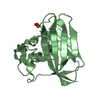
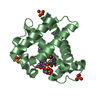
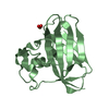
Neutron crystallographic structure of a Jonesia denitrificans lytic polysaccharide monooxygenase
ComponentsChitinase
KeywordsSUGAR BINDING PROTEIN / histidine brace / HYDROLASE
Function / homology
Function and homology information


chitinase / chitinase activity / chitin catabolic process / chitin binding / carbohydrate binding / carbohydrate metabolic process / extracellular region / metal ion binding
Similarity search - Function
Cellulose/chitin-binding protein, N-terminal / Lytic polysaccharide mono-oxygenase, cellulose-degrading / Carbohydrate-binding module family 5/12 / Chitin-binding domain type 3 / Carbohydrate-binding module family 5/12 / Carbohydrate-binding module superfamily 5/12 / Glycosyl hydrolases family 18 (GH18) active site / Glycosyl hydrolases family 18 (GH18) active site signature. / Chitinase insertion domain superfamily / Chitinase II ...Cellulose/chitin-binding protein, N-terminal / Lytic polysaccharide mono-oxygenase, cellulose-degrading / Carbohydrate-binding module family 5/12 / Chitin-binding domain type 3 / Carbohydrate-binding module family 5/12 / Carbohydrate-binding module superfamily 5/12 / Glycosyl hydrolases family 18 (GH18) active site / Glycosyl hydrolases family 18 (GH18) active site signature. / Chitinase insertion domain superfamily / Chitinase II / Glyco_18 / Glycosyl hydrolases family 18 (GH18) domain profile. / Glycoside hydrolase family 18, catalytic domain / Glycosyl hydrolases family 18 / Immunoglobulin E-set / Glycoside hydrolase superfamily
Similarity search - Domain/homology
COPPER (II) ION / PEROXIDE ION / Chitinase
Similarity search - Component
Biological speciesJonesia denitrificans (bacteria)
MethodNEUTRON DIFFRACTION / NUCLEAR REACTOR / MOLECULAR REPLACEMENT / Resolution: 2.1 Å
AuthorsBacik, J.-P. / Unkefer, C.J. / Chen, J.C.H.
CitationJournal: Biochemistry / Year: 2017
Title: Neutron and Atomic Resolution X-ray Structures of a Lytic Polysaccharide Monooxygenase Reveal Copper-Mediated Dioxygen Binding and Evidence for N-Terminal Deprotonation.
Authors: Bacik, J.P. / Mekasha, S. / Forsberg, Z. / Kovalevsky, A.Y. / Vaaje-Kolstad, G. / Eijsink, V.G.H. / Nix, J.C. / Coates, L. / Cuneo, M.J. / Unkefer, C.J. / Chen, J.C.
History
DepositionApr 10, 2017Deposition site: RCSB / Processing site: RCSB
Revision 1.0May 24, 2017Provider: repository / Type: Initial release
Revision 1.1May 31, 2017Group: Database references
Revision 1.2Oct 4, 2023Group: Data collection / Database references ...Data collection / Database references / Derived calculations / Refinement description
Category: chem_comp_atom / chem_comp_bond ...chem_comp_atom / chem_comp_bond / database_2 / diffrn_source / pdbx_initial_refinement_model / pdbx_struct_conn_angle / struct_conn
Item: _database_2.pdbx_DOI / _database_2.pdbx_database_accession ..._database_2.pdbx_DOI / _database_2.pdbx_database_accession / _diffrn_source.pdbx_synchrotron_beamline / _diffrn_source.pdbx_synchrotron_site / _pdbx_struct_conn_angle.ptnr1_auth_asym_id / _pdbx_struct_conn_angle.ptnr1_auth_comp_id / _pdbx_struct_conn_angle.ptnr1_auth_seq_id / _pdbx_struct_conn_angle.ptnr1_label_asym_id / _pdbx_struct_conn_angle.ptnr1_label_atom_id / _pdbx_struct_conn_angle.ptnr1_label_comp_id / _pdbx_struct_conn_angle.ptnr1_label_seq_id / _pdbx_struct_conn_angle.ptnr3_auth_asym_id / _pdbx_struct_conn_angle.ptnr3_auth_comp_id / _pdbx_struct_conn_angle.ptnr3_auth_seq_id / _pdbx_struct_conn_angle.ptnr3_label_asym_id / _pdbx_struct_conn_angle.ptnr3_label_atom_id / _pdbx_struct_conn_angle.ptnr3_label_comp_id / _pdbx_struct_conn_angle.ptnr3_label_seq_id / _pdbx_struct_conn_angle.value / _struct_conn.pdbx_dist_value / _struct_conn.ptnr1_auth_asym_id / _struct_conn.ptnr1_auth_comp_id / _struct_conn.ptnr1_auth_seq_id / _struct_conn.ptnr1_label_asym_id / _struct_conn.ptnr1_label_atom_id / _struct_conn.ptnr1_label_comp_id / _struct_conn.ptnr1_label_seq_id / _struct_conn.ptnr2_auth_asym_id / _struct_conn.ptnr2_auth_comp_id / _struct_conn.ptnr2_auth_seq_id / _struct_conn.ptnr2_label_asym_id / _struct_conn.ptnr2_label_atom_id / _struct_conn.ptnr2_label_comp_id

-
Structure visualization

Structure viewerMolecule:
MolmilJmol/JSmol

Downloads & links

-
Assembly

Deposited unit
A: Chitinase
B: Chitinase
hetero molecules


Theoretical massNumber of molelcules
Total (without water)31,2455
Polymers31,0862
Non-polymers1593
Water4,161231
1
A: Chitinase
hetero molecules


Theoretical massNumber of molelcules
Total (without water)15,6062
Polymers15,5431
Non-polymers641
Water181
TypeNameSymmetry operationNumber
identity operation1_555x,y,z1
MethodPISA
2
B: Chitinase
hetero molecules


Theoretical massNumber of molelcules
Total (without water)15,6383
Polymers15,5431
Non-polymers962
Water181
TypeNameSymmetry operationNumber
identity operation1_555x,y,z1
MethodPISA
Unit cell
Length a, b, c (Å)32.500, 76.400, 122.100
Angle α, β, γ (deg.)90.00, 90.00, 90.00
Int Tables number19
Space group name H-MP212121

-
Components

#1: Protein Chitinase


Mass: 15542.896 Da / Num. of mol.: 2 / Fragment: residues 32-173
Source method: isolated from a genetically manipulated source
Source: (gene. exp.) Jonesia denitrificans (strain ATCC 14870 / DSM 20603 / CIP 55134) (bacteria)
Strain: ATCC 14870 / DSM 20603 / CIP 55134 / Gene: Jden_1381 / Production host: Escherichia coli (E. coli) / References: UniProt: C7R4I0, chitinase
#2: Chemical ChemComp-CU / COPPER (II) ION


Mass: 63.546 Da / Num. of mol.: 2 / Source method: obtained synthetically / Formula: Cu
#3: Chemical ChemComp-PER / PEROXIDE ION


Mass: 31.999 Da / Num. of mol.: 1 / Source method: obtained synthetically / Formula: O2
#4: Water ChemComp-HOH / water


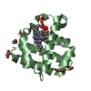
Mass: 18.015 Da / Num. of mol.: 231 / Source method: isolated from a natural source / Formula: H2O

-
Experimental details

-
Experiment

ExperimentMethod: NEUTRON DIFFRACTION / Number of used crystals: 1

-
Sample preparation

Crystal grow
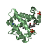
Temperature: 293 K / Method: vapor diffusion, sitting drop
Details: Purified enzyme was incubated with a threefold molar excess of CuSO4 for 30 min at room temperature. To remove excess copper, the protein was loaded onto a desalting column equilibrated with ...Details: Purified enzyme was incubated with a threefold molar excess of CuSO4 for 30 min at room temperature. To remove excess copper, the protein was loaded onto a desalting column equilibrated with 20 mM Tris-HCl pH 8.0. 14 ul protein solution at 48 mg/ml was mixed with 14 ul reservoir buffer consisting of 1.9 M DL-malic acid pH 7.0

-
Data collection

DiffractionMean temperature: 295 K
Diffraction sourceSource: NUCLEAR REACTOR / Site: ORNL Spallation Neutron Source / Beamline: MANDI / Wavelength: 2-4
DetectorType: ORNL ANGER CAMERA / Detector: AREA DETECTOR / Date: May 21, 2015
RadiationProtocol: LAUE / Monochromatic (M) / Laue (L): L / Scattering type: neutron
Radiation wavelength
IDWavelength (Å)Relative weight
121
241
ReflectionResolution: 2.1→12 Å / Num. obs: 13989 / % possible obs: 76 % / Redundancy: 4.5 % / Net I/σ(I): 9.4
Reflection shellResolution: 2.1→2.17 Å / Redundancy: 2.5 % / Mean I/σ(I) obs: 2.7 / % possible all: 59.4

-
Processing

Software
NameVersionClassification
PHENIX(1.10.1_2155)refinement
PHENIXphasing
Mantiddata reduction
RefinementMethod to determine structure: MOLECULAR REPLACEMENT
Starting model: 5VG0
Resolution: 2.1→12 Å / SU ML: 0.35 / Cross valid method: FREE R-VALUE / σ(F): 2.38 / Phase error: 25.82
RfactorNum. reflection% reflection
Rfree0.265 686 4.9 %
Rwork0.187 --
obs0.191 13989 76 %
Solvent computationShrinkage radii: 0.9 Å / VDW probe radii: 1.11 Å
Refine LS restraints
Refine-IDTypeDev idealNumber
NEUTRON DIFFRACTIONf_bond_d0.0025222
NEUTRON DIFFRACTIONf_angle_d0.568788
NEUTRON DIFFRACTIONf_dihedral_angle_d11.892390
NEUTRON DIFFRACTIONf_chiral_restr0.04350
NEUTRON DIFFRACTIONf_plane_restr0.0021025
LS refinement shell
Resolution (Å)Rfactor RfreeNum. reflection RfreeRfactor RworkNum. reflection RworkRefine-ID% reflection obs (%)
2.1001-2.26120.35181150.26292181NEUTRON DIFFRACTION64
2.2612-2.4870.32271390.23162544NEUTRON DIFFRACTION74
2.487-2.84270.29481360.21042781NEUTRON DIFFRACTION80
2.8427-3.56590.24641510.19262930NEUTRON DIFFRACTION83
3.5659-12.03290.22261450.14542867NEUTRON DIFFRACTION78

+
About Yorodumi

-
News

-
Feb 9, 2022. New format data for meta-information of EMDB entries

New format data for meta-information of EMDB entries

  • Version 3 of the EMDB header file is now the official format.
  • The previous official version 1.9 will be removed from the archive.

Related info.:EMDB header

External links:wwPDB to switch to version 3 of the EMDB data model

-
Aug 12, 2020. Covid-19 info

Covid-19 info

URL: https://pdbjlvh1.pdbj.org/emnavi/covid19.php

New page: Covid-19 featured information page in EM Navigator.

Related info.:Covid-19 info / Mar 5, 2020. Novel coronavirus structure data

+
Mar 5, 2020. Novel coronavirus structure data

Novel coronavirus structure data

Related info.:Yorodumi Speices / Aug 12, 2020. Covid-19 info

External links:COVID-19 featured content - PDBj / Molecule of the Month (242):Coronavirus Proteases

+
Jan 31, 2019. EMDB accession codes are about to change! (news from PDBe EMDB page)

EMDB accession codes are about to change! (news from PDBe EMDB page)

  • The allocation of 4 digits for EMDB accession codes will soon come to an end. Whilst these codes will remain in use, new EMDB accession codes will include an additional digit and will expand incrementally as the available range of codes is exhausted. The current 4-digit format prefixed with “EMD-” (i.e. EMD-XXXX) will advance to a 5-digit format (i.e. EMD-XXXXX), and so on. It is currently estimated that the 4-digit codes will be depleted around Spring 2019, at which point the 5-digit format will come into force.
  • The EM Navigator/Yorodumi systems omit the EMD- prefix.

Related info.:Q: What is EMD? / ID/Accession-code notation in Yorodumi/EM Navigator

External links:EMDB Accession Codes are Changing Soon! / Contact to PDBj

+
Jul 12, 2017. Major update of PDB

Major update of PDB

  • wwPDB released updated PDB data conforming to the new PDBx/mmCIF dictionary.
  • This is a major update changing the version number from 4 to 5, and with Remediation, in which all the entries are updated.
  • In this update, many items about electron microscopy experimental information are reorganized (e.g. em_software).
  • Now, EM Navigator and Yorodumi are based on the updated data.

External links:wwPDB Remediation / Enriched Model Files Conforming to OneDep Data Standards Now Available in the PDB FTP Archive

-
Yorodumi

Thousand views of thousand structures

  • Yorodumi is a browser for structure data from EMDB, PDB, SASBDB, etc.
  • This page is also the successor to EM Navigator detail page, and also detail information page/front-end page for Omokage search.
  • The word "yorodu" (or yorozu) is an old Japanese word meaning "ten thousand". "mi" (miru) is to see.

Related info.:EMDB / PDB / SASBDB / Comparison of 3 databanks / Yorodumi Search / Aug 31, 2016. New EM Navigator & Yorodumi / Yorodumi Papers / Jmol/JSmol / Function and homology information / Changes in new EM Navigator and Yorodumi

Read more