 Movie
Movie Controller


+ Open data

- Basic information
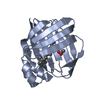
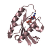
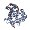
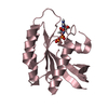
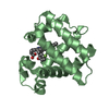
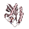
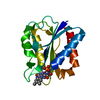
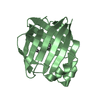
| Entry | Database: PDB / ID: 4kl5 | ||||||
|---|---|---|---|---|---|---|---|
| Title | Crystal structure of NpuDnaE intein | ||||||
 Components Components | DNA polymerase III, alpha subunit, Nucleic acid binding, OB-fold, tRNA/helicase-type chimeric construct | ||||||
 Keywords Keywords | UNKNOWN FUNCTION / HINT / intein / NpuDnaE inten | ||||||
| Function / homology |  Function and homology information Function and homology informationintein-mediated protein splicing / 3'-5' exonuclease activity / helicase activity / DNA-directed DNA polymerase / nucleic acid binding / DNA-directed DNA polymerase activity / DNA replication / metal ion binding Similarity search - Function | ||||||
| Biological species |  Nostoc punctiforme (bacteria) Nostoc punctiforme (bacteria) | ||||||
| Method |  X-RAY DIFFRACTION / SYNCHROTRON / MOLECULAR REPLACEMENT / Resolution: 1.72 Å X-RAY DIFFRACTION / SYNCHROTRON / MOLECULAR REPLACEMENT / Resolution: 1.72 Å | ||||||
 Authors Authors | Aranko, A.S. / Oeemig, J.S. / Kajander, T. / Iwai, H. | ||||||
 Citation Citation | Journal: Nat.Chem.Biol. / Year: 2013 Title: Intermolecular domain swapping induces intein-mediated protein alternative splicing. Authors: Aranko, A.S. / Oeemig, J.S. / Kajander, T. / Iwai, H. | ||||||
| History |
|
- Structure visualization
| Structure viewer | Molecule: MolmilJmol/JSmol |
|---|
- Downloads & links
-Download
| PDBx/mmCIF format | 4kl5.cif.gz | 138.1 KB | Display | PDBx/mmCIF format |
|---|---|---|---|---|
| PDB format | pdb4kl5.ent.gz | 107.5 KB | Display | PDB format |
| PDBx/mmJSON format | 4kl5.json.gz | Tree view | PDBx/mmJSON format | |
| Others |  Other downloads Other downloads |
-Validation report
| Arichive directory | https://data.pdbj.org/pub/pdb/validation_reports/kl/4kl5ftp://data.pdbj.org/pub/pdb/validation_reports/kl/4kl5 | HTTPS FTP |
|---|
-Related structure data
| Related structure data | 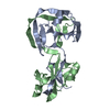 4kl6C 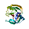 2keqS S: Starting model for refinement C: citing same article ( |
|---|---|
| Similar structure data |


-Links
 PDBj
PDBj


- Assembly
| Deposited unit | 
| ||||||||
|---|---|---|---|---|---|---|---|---|---|
| 1 | 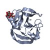
| ||||||||
| 2 | 
| ||||||||
| 3 | 
| ||||||||
| Unit cell |
|
-Components
| #1: Protein | Mass: 16377.375 Da / Num. of mol.: 2 Fragment: NpuDnaE Intein (unp residues 775-876, unp residues 2-40) Mutation: C1A, D142N, D143S Source method: isolated from a genetically manipulated source Source: (gene. exp.) Nostoc punctiforme (bacteria) / Strain: ATCC 29133 / PCC 73102 / Gene: DnaE N- and C-intein part, Npun_F4872 / Plasmid: pALBRSF12 / Production host: References: UniProt: B2J066, UniProt: B2J821, DNA-directed DNA polymerase #2: Chemical |   Mass: 192.124 Da / Num. of mol.: 2 / Source method: obtained synthetically / Formula: C6H8O7 Mass: 192.124 Da / Num. of mol.: 2 / Source method: obtained synthetically / Formula: C6H8O7#3: Chemical | ChemComp-GOL / |   Mass: 92.094 Da / Num. of mol.: 1 / Source method: obtained synthetically / Formula: C3H8O3 Mass: 92.094 Da / Num. of mol.: 1 / Source method: obtained synthetically / Formula: C3H8O3#4: Water | ChemComp-HOH / |  Mass: 18.015 Da / Num. of mol.: 221 / Source method: isolated from a natural source / Formula: H2O Mass: 18.015 Da / Num. of mol.: 221 / Source method: isolated from a natural source / Formula: H2O |
|---|
-Experimental details
-Experiment
| Experiment | Method: X-RAY DIFFRACTION / Number of used crystals: 1 |
|---|
- Sample preparation
| Crystal | Density Matthews: 1.98 Å3/Da / Density % sol: 37.8 % |
|---|---|
| Crystal grow | Temperature: 293 K / Method: vapor diffusion, sitting drop / pH: 6.5 Details: 1.4 M tri-ammonium citrate/citric acid , pH 6.5, VAPOR DIFFUSION, SITTING DROP, temperature 293K |
-Data collection
| Diffraction | Mean temperature: 100 K | ||||||||||||||||||||||||||||||||||||||||||||||||||||||||||||||||||||||
|---|---|---|---|---|---|---|---|---|---|---|---|---|---|---|---|---|---|---|---|---|---|---|---|---|---|---|---|---|---|---|---|---|---|---|---|---|---|---|---|---|---|---|---|---|---|---|---|---|---|---|---|---|---|---|---|---|---|---|---|---|---|---|---|---|---|---|---|---|---|---|---|
| Diffraction source | Source: SYNCHROTRON / Site: ESRF  / Beamline: ID23-1 / Wavelength: 0.976 Å / Beamline: ID23-1 / Wavelength: 0.976 Å | ||||||||||||||||||||||||||||||||||||||||||||||||||||||||||||||||||||||
| Detector | Type: ADSC QUANTUM 315r / Detector: CCD / Date: Feb 6, 2011 | ||||||||||||||||||||||||||||||||||||||||||||||||||||||||||||||||||||||
| Radiation | Monochromator: Si(111) crystal / Protocol: SINGLE WAVELENGTH / Monochromatic (M) / Laue (L): M / Scattering type: x-ray | ||||||||||||||||||||||||||||||||||||||||||||||||||||||||||||||||||||||
| Radiation wavelength | Wavelength: 0.976 Å / Relative weight: 1 | ||||||||||||||||||||||||||||||||||||||||||||||||||||||||||||||||||||||
| Reflection | Resolution: 1.72→50 Å / Num. all: 28489 / Num. obs: 28252 / % possible obs: 99.2 % / Observed criterion σ(F): 0 / Observed criterion σ(I): -3 / Redundancy: 7.04 % / Rmerge(I) obs: 0.065 | ||||||||||||||||||||||||||||||||||||||||||||||||||||||||||||||||||||||
| Reflection shell |
|
- Processing
| Software |
| |||||||||||||||||||||||||||||||||||||||||||||||||||||||||||||||||||||||||||||
|---|---|---|---|---|---|---|---|---|---|---|---|---|---|---|---|---|---|---|---|---|---|---|---|---|---|---|---|---|---|---|---|---|---|---|---|---|---|---|---|---|---|---|---|---|---|---|---|---|---|---|---|---|---|---|---|---|---|---|---|---|---|---|---|---|---|---|---|---|---|---|---|---|---|---|---|---|---|---|
| Refinement | Method to determine structure: MOLECULAR REPLACEMENT Starting model: PDB entry 2KEQ Resolution: 1.72→47.439 Å / SU ML: 0.16 / σ(F): 3.67 / σ(I): 3.67 / Phase error: 19.97 / Stereochemistry target values: MLHL
| |||||||||||||||||||||||||||||||||||||||||||||||||||||||||||||||||||||||||||||
| Solvent computation | Shrinkage radii: 0.9 Å / VDW probe radii: 1.11 Å / Solvent model: FLAT BULK SOLVENT MODEL | |||||||||||||||||||||||||||||||||||||||||||||||||||||||||||||||||||||||||||||
| Refinement step | Cycle: LAST / Resolution: 1.72→47.439 Å
| |||||||||||||||||||||||||||||||||||||||||||||||||||||||||||||||||||||||||||||
| Refine LS restraints |
| |||||||||||||||||||||||||||||||||||||||||||||||||||||||||||||||||||||||||||||
| LS refinement shell |
| |||||||||||||||||||||||||||||||||||||||||||||||||||||||||||||||||||||||||||||
| Refinement TLS params. | Method: refined / Origin x: 1.4747 Å / Origin y: 1.2687 Å / Origin z: 8.8625 Å
| |||||||||||||||||||||||||||||||||||||||||||||||||||||||||||||||||||||||||||||
| Refinement TLS group | Selection details: all |
