- PDB-5aa7: Structural and functional characterization of a chitin-active 15.... -
+
Open data
ID or keywords:
Loading...
-
Basic information
Entry
Database: PDB / ID: 5aa7
Title
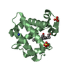
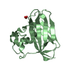
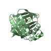
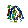
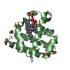
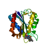
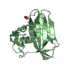
Structural and functional characterization of a chitin-active 15.5 kDa lytic polysaccharide monooxygenase domain from a modular chitinase from Jonesia denitrificans
Components
CHITINASE
Keywords
HYDROLASE
Function / homology
Function and homology information
endochitinase activity / chitinase / chitin catabolic process / chitin binding / carbohydrate binding / carbohydrate metabolic process / extracellular region / metal ion binding Similarity search - Function
chitin-binding protein cbp21 / Cellulose/chitin-binding protein, N-terminal / Lytic polysaccharide mono-oxygenase, cellulose-degrading / Carbohydrate-binding module family 5/12 / Chitin-binding domain type 3 / Carbohydrate-binding module family 5/12 / Carbohydrate-binding module superfamily 5/12 / Coagulation Factor XIII; Chain A, domain 1 / Glycosyl hydrolases family 18 (GH18) active site / Glycosyl hydrolases family 18 (GH18) active site signature. ...chitin-binding protein cbp21 / Cellulose/chitin-binding protein, N-terminal / Lytic polysaccharide mono-oxygenase, cellulose-degrading / Carbohydrate-binding module family 5/12 / Chitin-binding domain type 3 / Carbohydrate-binding module family 5/12 / Carbohydrate-binding module superfamily 5/12 / Coagulation Factor XIII; Chain A, domain 1 / Glycosyl hydrolases family 18 (GH18) active site / Glycosyl hydrolases family 18 (GH18) active site signature. / : / Chitinase insertion domain superfamily / Chitinase II / Glyco_18 / Glycosyl hydrolases family 18 / Glycosyl hydrolases family 18 (GH18) domain profile. / Glycoside hydrolase family 18, catalytic domain / Distorted Sandwich / Immunoglobulin E-set / Glycoside hydrolase superfamily / Mainly Beta Similarity search - Domain/homology
In the structure databanks used in Yorodumi, some data are registered as the other names, "COVID-19 virus" and "2019-nCoV". Here are the details of the virus and the list of structure data.
Jan 31, 2019. EMDB accession codes are about to change! (news from PDBe EMDB page)
EMDB accession codes are about to change! (news from PDBe EMDB page)
The allocation of 4 digits for EMDB accession codes will soon come to an end. Whilst these codes will remain in use, new EMDB accession codes will include an additional digit and will expand incrementally as the available range of codes is exhausted. The current 4-digit format prefixed with “EMD-” (i.e. EMD-XXXX) will advance to a 5-digit format (i.e. EMD-XXXXX), and so on. It is currently estimated that the 4-digit codes will be depleted around Spring 2019, at which point the 5-digit format will come into force.
The EM Navigator/Yorodumi systems omit the EMD- prefix.
Related info.:Q: What is EMD? / ID/Accession-code notation in Yorodumi/EM Navigator
Yorodumi is a browser for structure data from EMDB, PDB, SASBDB, etc.
This page is also the successor to EM Navigator detail page, and also detail information page/front-end page for Omokage search.
The word "yorodu" (or yorozu) is an old Japanese word meaning "ten thousand". "mi" (miru) is to see.
Related info.:EMDB / PDB / SASBDB / Comparison of 3 databanks / Yorodumi Search / Aug 31, 2016. New EM Navigator & Yorodumi / Yorodumi Papers / Jmol/JSmol / Function and homology information / Changes in new EM Navigator and Yorodumi
 Movie
Movie Controller

 Yorodumi
 Open data
 Basic information
 Components
 Keywords
 Function and homology information
 JONESIA DENITRIFICANS (bacteria)
 X-RAY DIFFRACTION /
  Authors
 Citation
 Structure visualization
 Downloads & links
 Other downloads
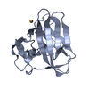


 PDBj
PDBj Assembly


 Mass: 63.546 Da / Num. of mol.: 2 / Source method: obtained synthetically / Formula: Cu
 Mass: 18.015 Da / Num. of mol.: 328 / Source method: isolated from a natural source / Formula: H2O
 Sample preparation
 / Beamline: ID29 / Wavelength: 1.37778
  Processing