 Movie
Movie Controller

[English] 日本語
 Yorodumi
- PDB-5vg0: Room temperature X-ray crystallographic structure of a Jonesia de... -
+ Open data

- Basic information
| Entry | Database: PDB / ID: 5vg0 | ||||||
|---|---|---|---|---|---|---|---|
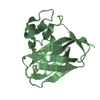
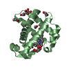
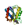
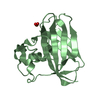
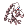
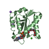
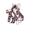
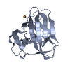
| Title | Room temperature X-ray crystallographic structure of a Jonesia denitrificans lytic polysaccharide monooxygenase at 1.1 angstrom resolution. | ||||||
 Components Components | Chitinase | ||||||
 Keywords Keywords | SUGAR BINDING PROTEIN / histidine brace / HYDROLASE | ||||||
| Function / homology |  Function and homology information Function and homology informationendochitinase activity / chitinase / chitin catabolic process / chitin binding / carbohydrate binding / carbohydrate metabolic process / extracellular region / metal ion binding Similarity search - Function | ||||||
| Biological species |  Jonesia denitrificans (bacteria) Jonesia denitrificans (bacteria) | ||||||
| Method |  X-RAY DIFFRACTION / SYNCHROTRON / MOLECULAR REPLACEMENT / Resolution: 1.1 Å X-RAY DIFFRACTION / SYNCHROTRON / MOLECULAR REPLACEMENT / Resolution: 1.1 Å | ||||||
 Authors Authors | Bacik, J.-P. / Unkefer, C.J. / Chen, J.C.H. | ||||||
 Citation Citation | Journal: Biochemistry / Year: 2017 Title: Neutron and Atomic Resolution X-ray Structures of a Lytic Polysaccharide Monooxygenase Reveal Copper-Mediated Dioxygen Binding and Evidence for N-Terminal Deprotonation. Authors: Bacik, J.P. / Mekasha, S. / Forsberg, Z. / Kovalevsky, A.Y. / Vaaje-Kolstad, G. / Eijsink, V.G.H. / Nix, J.C. / Coates, L. / Cuneo, M.J. / Unkefer, C.J. / Chen, J.C. | ||||||
| History |
|
- Structure visualization
| Structure viewer | Molecule: MolmilJmol/JSmol |
|---|
- Downloads & links
-Download
| PDBx/mmCIF format | 5vg0.cif.gz | 188.1 KB | Display | PDBx/mmCIF format |
|---|---|---|---|---|
| PDB format | pdb5vg0.ent.gz | 153.7 KB | Display | PDB format |
| PDBx/mmJSON format | 5vg0.json.gz | Tree view | PDBx/mmJSON format | |
| Others |  Other downloads Other downloads |
-Validation report
| Arichive directory | https://data.pdbj.org/pub/pdb/validation_reports/vg/5vg0ftp://data.pdbj.org/pub/pdb/validation_reports/vg/5vg0 | HTTPS FTP |
|---|
-Related structure data
| Related structure data |  5vg1C  5aa7S C: citing same article ( S: Starting model for refinement |
|---|---|
| Similar structure data |


-Links
 PDBj
PDBj- Assembly
| Deposited unit | 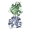
| ||||||||
|---|---|---|---|---|---|---|---|---|---|
| 1 | 
| ||||||||
| 2 | 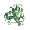
| ||||||||
| Unit cell |
|
-Components
| #1: Protein | Mass: 15542.896 Da / Num. of mol.: 2 / Fragment: residues 32-173 Source method: isolated from a genetically manipulated source Source: (gene. exp.) Jonesia denitrificans (strain ATCC 14870 / DSM 20603 / CIP 55134) (bacteria)Strain: ATCC 14870 / DSM 20603 / CIP 55134 / Gene: Jden_1381 / Production host: #2: Chemical |   Mass: 63.546 Da / Num. of mol.: 2 / Source method: obtained synthetically / Formula: Cu Mass: 63.546 Da / Num. of mol.: 2 / Source method: obtained synthetically / Formula: Cu#3: Chemical |   Mass: 31.999 Da / Num. of mol.: 2 / Source method: obtained synthetically / Formula: O2 Mass: 31.999 Da / Num. of mol.: 2 / Source method: obtained synthetically / Formula: O2#4: Water | ChemComp-HOH / |  Mass: 18.015 Da / Num. of mol.: 299 / Source method: isolated from a natural source / Formula: H2O Mass: 18.015 Da / Num. of mol.: 299 / Source method: isolated from a natural source / Formula: H2OHas protein modification | Y | |
|---|
-Experimental details
-Experiment
| Experiment | Method: X-RAY DIFFRACTION / Number of used crystals: 1 |
|---|
- Sample preparation
| Crystal | Density Matthews: 2.34 Å3/Da / Density % sol: 47.51 % |
|---|---|
| Crystal grow | Temperature: 293 K / Method: vapor diffusion, sitting drop Details: Purified enzyme was incubated with a threefold molar excess of CuSO4 for 30 min at room temperature. To remove excess copper, the protein was loaded onto a desalting column equilibrated with ...Details: Purified enzyme was incubated with a threefold molar excess of CuSO4 for 30 min at room temperature. To remove excess copper, the protein was loaded onto a desalting column equilibrated with 20 mM Tris-HCl pH 8.0. 30 ul protein solution at 48 mg/ml was mixed with 30 ul reservoir buffer consisting of 1.9 M DL-malic acid pH 7.0 |
-Data collection
| Diffraction | Mean temperature: 295 K |
|---|---|
| Diffraction source | Source: SYNCHROTRON / Site: ALS  / Beamline: 4.2.2 / Wavelength: 1 Å / Beamline: 4.2.2 / Wavelength: 1 Å |
| Detector | Type: RDI CMOS_8M / Detector: CMOS / Date: May 27, 2015 |
| Radiation | Protocol: SINGLE WAVELENGTH / Monochromatic (M) / Laue (L): M / Scattering type: x-ray |
| Radiation wavelength | Wavelength: 1 Å / Relative weight: 1 |
| Reflection | Resolution: 1.1→32 Å / Num. obs: 1248110 / % possible obs: 97.3 % / Redundancy: 10.7 % / Net I/σ(I): 13.7 |
| Reflection shell | Resolution: 1.1→1.16 Å / Redundancy: 10.4 % / % possible all: 95 |
- Processing
| Software |
| |||||||||||||||||||||||||||||||||||||||||||||||||||||||||||||||||||||||||||||||||||||||||||||||||||||||||
|---|---|---|---|---|---|---|---|---|---|---|---|---|---|---|---|---|---|---|---|---|---|---|---|---|---|---|---|---|---|---|---|---|---|---|---|---|---|---|---|---|---|---|---|---|---|---|---|---|---|---|---|---|---|---|---|---|---|---|---|---|---|---|---|---|---|---|---|---|---|---|---|---|---|---|---|---|---|---|---|---|---|---|---|---|---|---|---|---|---|---|---|---|---|---|---|---|---|---|---|---|---|---|---|---|---|---|
| Refinement | Method to determine structure: MOLECULAR REPLACEMENT Starting model: 5AA7 Resolution: 1.1→32 Å / SU ML: 0.08 / Cross valid method: FREE R-VALUE / σ(F): 1.33 / Phase error: 13.51
| |||||||||||||||||||||||||||||||||||||||||||||||||||||||||||||||||||||||||||||||||||||||||||||||||||||||||
| Solvent computation | Shrinkage radii: 0.9 Å / VDW probe radii: 1.11 Å | |||||||||||||||||||||||||||||||||||||||||||||||||||||||||||||||||||||||||||||||||||||||||||||||||||||||||
| Refinement step | Cycle: LAST / Resolution: 1.1→32 Å
| |||||||||||||||||||||||||||||||||||||||||||||||||||||||||||||||||||||||||||||||||||||||||||||||||||||||||
| Refine LS restraints |
| |||||||||||||||||||||||||||||||||||||||||||||||||||||||||||||||||||||||||||||||||||||||||||||||||||||||||
| LS refinement shell |
|
