 Movie
Movie Controller

[English] 日本語
 Yorodumi
- PDB-5vc6: crystal structure of human WEE1 kinase domain in complex with PHA... -
+ Open data

- Basic information
| Entry | Database: PDB / ID: 5vc6 | ||||||
|---|---|---|---|---|---|---|---|
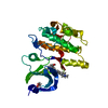
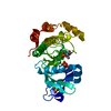
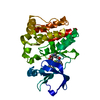
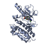
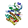
| Title | crystal structure of human WEE1 kinase domain in complex with PHA-848125 | ||||||
 Components Components | Wee1-like protein kinase | ||||||
 Keywords Keywords | TRANSFERASE/TRANSFERASE INHIBITOR / KINASE DOMAIN / CELL CYCLE / WEE1 / TRANSFERASE / INHIBITOR / TRANSFERASE-TRANSFERASE INHIBITOR complex | ||||||
| Function / homology |  Function and homology information Function and homology informationnegative regulation of G2/M transition of mitotic cell cycle / G2/M DNA replication checkpoint / Polo-like kinase mediated events / negative regulation of G1/S transition of mitotic cell cycle / establishment of cell polarity / Cyclin E associated events during G1/S transition / Cyclin A:Cdk2-associated events at S phase entry / Cyclin A/B1/B2 associated events during G2/M transition / Chk1/Chk2(Cds1) mediated inactivation of Cyclin B:Cdk1 complex / neuron projection morphogenesis ...negative regulation of G2/M transition of mitotic cell cycle / G2/M DNA replication checkpoint / Polo-like kinase mediated events / negative regulation of G1/S transition of mitotic cell cycle / establishment of cell polarity / Cyclin E associated events during G1/S transition / Cyclin A:Cdk2-associated events at S phase entry / Cyclin A/B1/B2 associated events during G2/M transition / Chk1/Chk2(Cds1) mediated inactivation of Cyclin B:Cdk1 complex / neuron projection morphogenesis / positive regulation of DNA replication / non-specific protein-tyrosine kinase / non-membrane spanning protein tyrosine kinase activity / microtubule cytoskeleton organization / G2/M transition of mitotic cell cycle / Factors involved in megakaryocyte development and platelet production / protein tyrosine kinase activity / cell division / nucleolus / magnesium ion binding / nucleoplasm / ATP binding / nucleus / cytoplasm Similarity search - Function | ||||||
| Biological species |  Homo sapiens (human) Homo sapiens (human) | ||||||
| Method |  X-RAY DIFFRACTION / MOLECULAR REPLACEMENT / molecular replacement / Resolution: 2 Å X-RAY DIFFRACTION / MOLECULAR REPLACEMENT / molecular replacement / Resolution: 2 Å | ||||||
 Authors Authors | Zhu, J.-Y. / Schonbrunn, E. | ||||||
| Funding support |  United States, 1items United States, 1items
| ||||||
 Citation Citation | Journal: J. Med. Chem. / Year: 2017 Title: Structural Basis of Wee Kinases Functionality and Inactivation by Diverse Small Molecule Inhibitors. Authors: Zhu, J.Y. / Cuellar, R.A. / Berndt, N. / Lee, H.E. / Olesen, S.H. / Martin, M.P. / Jensen, J.T. / Georg, G.I. / Schonbrunn, E. | ||||||
| History |
|
- Structure visualization
| Structure viewer | Molecule: MolmilJmol/JSmol |
|---|
- Downloads & links
-Download
| PDBx/mmCIF format | 5vc6.cif.gz | 168.1 KB | Display | PDBx/mmCIF format |
|---|---|---|---|---|
| PDB format | pdb5vc6.ent.gz | 133.3 KB | Display | PDB format |
| PDBx/mmJSON format | 5vc6.json.gz | Tree view | PDBx/mmJSON format | |
| Others |  Other downloads Other downloads |
-Validation report
| Arichive directory | https://data.pdbj.org/pub/pdb/validation_reports/vc/5vc6ftp://data.pdbj.org/pub/pdb/validation_reports/vc/5vc6 | HTTPS FTP |
|---|
-Related structure data
| Related structure data |  5v5ySC  5vc3C  5vc4C  5vc5C  5vcvC  5vcwC  5vcxC  5vcyC  5vczC  5vd0C  5vd1C  5vd2C  5vd3C  5vdkC C: citing same article ( S: Starting model for refinement |
|---|---|
| Similar structure data |


-Links
 PDBj
PDBj


- Assembly
| Deposited unit | 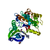
| ||||||||
|---|---|---|---|---|---|---|---|---|---|
| 1 |
| ||||||||
| Unit cell |
|
-Components
| #1: Protein | Mass: 32440.900 Da / Num. of mol.: 1 / Fragment: UNP RESIDUES 291-575 Source method: isolated from a genetically manipulated source Source: (gene. exp.) Homo sapiens (human) / Gene: WEE1 / Plasmid: pET28a / Production host:  References: UniProt: P30291, non-specific protein-tyrosine kinase |
|---|---|
| #2: Chemical | ChemComp-P48 /   Mass: 460.575 Da / Num. of mol.: 1 / Source method: obtained synthetically / Formula: C25H32N8O Mass: 460.575 Da / Num. of mol.: 1 / Source method: obtained synthetically / Formula: C25H32N8O |
| #3: Chemical | ChemComp-EDO /   Mass: 62.068 Da / Num. of mol.: 1 / Source method: obtained synthetically / Formula: C2H6O2 Mass: 62.068 Da / Num. of mol.: 1 / Source method: obtained synthetically / Formula: C2H6O2 |
| #4: Water | ChemComp-HOH /  Mass: 18.015 Da / Num. of mol.: 94 / Source method: isolated from a natural source / Formula: H2O Mass: 18.015 Da / Num. of mol.: 94 / Source method: isolated from a natural source / Formula: H2O |
-Experimental details
-Experiment
| Experiment | Method: X-RAY DIFFRACTION / Number of used crystals: 1 |
|---|
- Sample preparation
| Crystal | Density Matthews: 2.21 Å3/Da / Density % sol: 44.22 % |
|---|---|
| Crystal grow | Temperature: 291 K / Method: vapor diffusion, hanging drop / pH: 6.9 Details: 5.0 MG/ML WEE1, 25 mM Na/K phosphate, 1 mM DTT, 0.05 M ammonium sulfate, 0.05 M Bis-tris (pH 5.5), 7.5 % PEG 3350, 1 mM PHA-848125 |
-Data collection
| Diffraction | Mean temperature: 93 K | ||||||||||||||||||||||||||||||||||||||||||||||||||||||||||||||||||||||||||||||||||||||||||||||||||||||||||||||||||||||||||||||||||||||||||||||||||||||||||||||||||||||||
|---|---|---|---|---|---|---|---|---|---|---|---|---|---|---|---|---|---|---|---|---|---|---|---|---|---|---|---|---|---|---|---|---|---|---|---|---|---|---|---|---|---|---|---|---|---|---|---|---|---|---|---|---|---|---|---|---|---|---|---|---|---|---|---|---|---|---|---|---|---|---|---|---|---|---|---|---|---|---|---|---|---|---|---|---|---|---|---|---|---|---|---|---|---|---|---|---|---|---|---|---|---|---|---|---|---|---|---|---|---|---|---|---|---|---|---|---|---|---|---|---|---|---|---|---|---|---|---|---|---|---|---|---|---|---|---|---|---|---|---|---|---|---|---|---|---|---|---|---|---|---|---|---|---|---|---|---|---|---|---|---|---|---|---|---|---|---|---|---|---|
| Diffraction source | Source: ROTATING ANODE / Type: RIGAKU MICROMAX-007 HF / Wavelength: 1.54178 Å | ||||||||||||||||||||||||||||||||||||||||||||||||||||||||||||||||||||||||||||||||||||||||||||||||||||||||||||||||||||||||||||||||||||||||||||||||||||||||||||||||||||||||
| Detector | Type: RIGAKU SATURN 944+ / Detector: CCD / Date: Nov 10, 2016 / Details: mirrors | ||||||||||||||||||||||||||||||||||||||||||||||||||||||||||||||||||||||||||||||||||||||||||||||||||||||||||||||||||||||||||||||||||||||||||||||||||||||||||||||||||||||||
| Radiation | Monochromator: mirrors / Protocol: SINGLE WAVELENGTH / Monochromatic (M) / Laue (L): M / Scattering type: x-ray | ||||||||||||||||||||||||||||||||||||||||||||||||||||||||||||||||||||||||||||||||||||||||||||||||||||||||||||||||||||||||||||||||||||||||||||||||||||||||||||||||||||||||
| Radiation wavelength | Wavelength: 1.54178 Å / Relative weight: 1 | ||||||||||||||||||||||||||||||||||||||||||||||||||||||||||||||||||||||||||||||||||||||||||||||||||||||||||||||||||||||||||||||||||||||||||||||||||||||||||||||||||||||||
| Reflection | Resolution: 2→63.163 Å / Num. obs: 18665 / % possible obs: 96.3 % / Observed criterion σ(I): -3 / Redundancy: 3.716 % / Biso Wilson estimate: 25.49 Å2 / CC1/2: 0.999 / Rmerge(I) obs: 0.045 / Rrim(I) all: 0.053 / Χ2: 0.935 / Net I/σ(I): 20.2 | ||||||||||||||||||||||||||||||||||||||||||||||||||||||||||||||||||||||||||||||||||||||||||||||||||||||||||||||||||||||||||||||||||||||||||||||||||||||||||||||||||||||||
| Reflection shell | Diffraction-ID: 1
|
-Phasing
| Phasing | Method: molecular replacement |
|---|
- Processing
| Software |
| ||||||||||||||||||||||||||||||||||||||||||||||||||||||||||||||||||||||||||||||||||||||||||||||||||||||||||||||||||||||||||||||||||||||||||||||||||||||
|---|---|---|---|---|---|---|---|---|---|---|---|---|---|---|---|---|---|---|---|---|---|---|---|---|---|---|---|---|---|---|---|---|---|---|---|---|---|---|---|---|---|---|---|---|---|---|---|---|---|---|---|---|---|---|---|---|---|---|---|---|---|---|---|---|---|---|---|---|---|---|---|---|---|---|---|---|---|---|---|---|---|---|---|---|---|---|---|---|---|---|---|---|---|---|---|---|---|---|---|---|---|---|---|---|---|---|---|---|---|---|---|---|---|---|---|---|---|---|---|---|---|---|---|---|---|---|---|---|---|---|---|---|---|---|---|---|---|---|---|---|---|---|---|---|---|---|---|---|---|---|---|
| Refinement | Method to determine structure: MOLECULAR REPLACEMENT Starting model: 5V5Y Resolution: 2→63.163 Å / SU ML: 0.28 / Cross valid method: FREE R-VALUE / σ(F): 1.37 / Phase error: 27.41
| ||||||||||||||||||||||||||||||||||||||||||||||||||||||||||||||||||||||||||||||||||||||||||||||||||||||||||||||||||||||||||||||||||||||||||||||||||||||
| Solvent computation | Shrinkage radii: 0.9 Å / VDW probe radii: 1.11 Å | ||||||||||||||||||||||||||||||||||||||||||||||||||||||||||||||||||||||||||||||||||||||||||||||||||||||||||||||||||||||||||||||||||||||||||||||||||||||
| Displacement parameters | Biso max: 107.76 Å2 / Biso mean: 37.6689 Å2 / Biso min: 11.6 Å2 | ||||||||||||||||||||||||||||||||||||||||||||||||||||||||||||||||||||||||||||||||||||||||||||||||||||||||||||||||||||||||||||||||||||||||||||||||||||||
| Refinement step | Cycle: final / Resolution: 2→63.163 Å
| ||||||||||||||||||||||||||||||||||||||||||||||||||||||||||||||||||||||||||||||||||||||||||||||||||||||||||||||||||||||||||||||||||||||||||||||||||||||
| Refine LS restraints |
| ||||||||||||||||||||||||||||||||||||||||||||||||||||||||||||||||||||||||||||||||||||||||||||||||||||||||||||||||||||||||||||||||||||||||||||||||||||||
| LS refinement shell | Refine-ID: X-RAY DIFFRACTION / Rfactor Rfree error: 0 / Total num. of bins used: 7
| ||||||||||||||||||||||||||||||||||||||||||||||||||||||||||||||||||||||||||||||||||||||||||||||||||||||||||||||||||||||||||||||||||||||||||||||||||||||
| Refinement TLS params. | Method: refined / Refine-ID: X-RAY DIFFRACTION
| ||||||||||||||||||||||||||||||||||||||||||||||||||||||||||||||||||||||||||||||||||||||||||||||||||||||||||||||||||||||||||||||||||||||||||||||||||||||
| Refinement TLS group |
|
