 Movie
Movie Controller

[English] 日本語
 Yorodumi
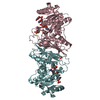
- PDB-5uib: Crystal Structure of an Oxidoreductase from Agrobacterium radioba... -
+ Open data

- Basic information
| Entry | Database: PDB / ID: 5uib | ||||||
|---|---|---|---|---|---|---|---|
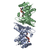
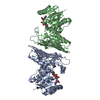
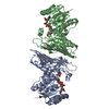
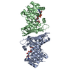
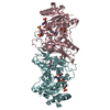
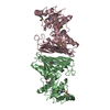
| Title | Crystal Structure of an Oxidoreductase from Agrobacterium radiobacter in Complex with NAD+, L-tartaric acid and Magnesium | ||||||
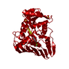
 Components Components | Oxidoreductase protein | ||||||
 Keywords Keywords | OXIDOREDUCTASE / L-tartrate | ||||||
| Function / homology |  Function and homology information Function and homology informationD-apiose dehydrogenase / oxidoreductase activity / nucleotide binding / metal ion binding Similarity search - Function | ||||||
| Biological species |  Agrobacterium radiobacter (Agrobacterium genomosp. 4) Agrobacterium radiobacter (Agrobacterium genomosp. 4) | ||||||
| Method |  X-RAY DIFFRACTION / SYNCHROTRON / MOLECULAR REPLACEMENT / Resolution: 2.65 Å X-RAY DIFFRACTION / SYNCHROTRON / MOLECULAR REPLACEMENT / Resolution: 2.65 Å | ||||||
 Authors Authors | Cook, W.J. / Fedorov, A.A. / Fedorov, E.V. / Huang, H. / Bonanno, J.B. / Gerlt, J.A. / Almo, S.C. | ||||||
| Funding support |  United States, 1items United States, 1items
| ||||||
 Citation Citation | Journal: To be published Title: Crystal Structure of an Oxidoreductase from Agrobacterium radiobacter in Complex with NAD+, L-tartaric acid and Magnesium Authors: Cook, W.J. / Fedorov, A.A. / Fedorov, E.V. / Huang, H. / Bonanno, J.B. / Gerlt, J.A. / Almo, S.C. | ||||||
| History |
|
- Structure visualization
| Structure viewer | Molecule: MolmilJmol/JSmol |
|---|
- Downloads & links
-Download
| PDBx/mmCIF format | 5uib.cif.gz | 270.8 KB | Display | PDBx/mmCIF format |
|---|---|---|---|---|
| PDB format | pdb5uib.ent.gz | 220.2 KB | Display | PDB format |
| PDBx/mmJSON format | 5uib.json.gz | Tree view | PDBx/mmJSON format | |
| Others |  Other downloads Other downloads |
-Validation report
| Arichive directory | https://data.pdbj.org/pub/pdb/validation_reports/ui/5uibftp://data.pdbj.org/pub/pdb/validation_reports/ui/5uib | HTTPS FTP |
|---|
-Related structure data
| Related structure data |  5uhwS S: Starting model for refinement |
|---|---|
| Similar structure data |


-Links
 PDBj
PDBj
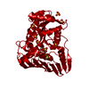
- Assembly
| Deposited unit | 
| ||||||||
|---|---|---|---|---|---|---|---|---|---|
| 1 |
| ||||||||
| Unit cell |
|
-Components
| #1: Protein | Mass: 37487.457 Da / Num. of mol.: 2 Source method: isolated from a genetically manipulated source Source: (gene. exp.) Agrobacterium radiobacter (strain K84 / ATCC BAA-868) (bacteria)Strain: K84 / ATCC BAA-868 / Gene: Arad_9238 / Plasmid: pET28a / Production host: #2: Chemical |   Mass: 663.425 Da / Num. of mol.: 2 / Source method: obtained synthetically / Formula: C21H27N7O14P2 / Comment: NAD*YM Mass: 663.425 Da / Num. of mol.: 2 / Source method: obtained synthetically / Formula: C21H27N7O14P2 / Comment: NAD*YM#3: Chemical |   Mass: 150.087 Da / Num. of mol.: 2 / Source method: obtained synthetically / Formula: C4H6O6 Mass: 150.087 Da / Num. of mol.: 2 / Source method: obtained synthetically / Formula: C4H6O6#4: Chemical |   Mass: 24.305 Da / Num. of mol.: 2 / Source method: obtained synthetically / Formula: Mg Mass: 24.305 Da / Num. of mol.: 2 / Source method: obtained synthetically / Formula: Mg#5: Water | ChemComp-HOH / |  Mass: 18.015 Da / Num. of mol.: 22 / Source method: isolated from a natural source / Formula: H2O Mass: 18.015 Da / Num. of mol.: 22 / Source method: isolated from a natural source / Formula: H2O |
|---|
-Experimental details
-Experiment
| Experiment | Method: X-RAY DIFFRACTION / Number of used crystals: 1 |
|---|
- Sample preparation
| Crystal | Density Matthews: 3.44 Å3/Da / Density % sol: 64.26 % |
|---|---|
| Crystal grow | Temperature: 293 K / Method: vapor diffusion, sitting drop / pH: 7 Details: 3.5 M Sodium formate, 5 mM NAD+, 5 mM magnesium chloride |
-Data collection
| Diffraction | Mean temperature: 100 K |
|---|---|
| Diffraction source | Source: SYNCHROTRON / Site: APS / Beamline: 31-ID / Wavelength: 0.97931 Å |
| Detector | Type: ADSC QUANTUM 315 / Detector: CCD / Date: Oct 19, 2016 / Details: Diamond |
| Radiation | Protocol: SINGLE WAVELENGTH / Monochromatic (M) / Laue (L): M / Scattering type: x-ray |
| Radiation wavelength | Wavelength: 0.97931 Å / Relative weight: 1 |
| Reflection | Resolution: 2.65→29.6 Å / Num. obs: 29362 / % possible obs: 99.4 % / Redundancy: 3.8 % / CC1/2: 0.994 / Rmerge(I) obs: 0.099 / Rpim(I) all: 0.094 / Rrim(I) all: 0.137 / Net I/σ(I): 5.5 |
| Reflection shell | Resolution: 2.65→2.79 Å / Redundancy: 3.8 % / Rmerge(I) obs: 0.827 / Mean I/σ(I) obs: 1.1 / Num. unique obs: 4299 / CC1/2: 0.538 / Rpim(I) all: 0.772 / Rrim(I) all: 1.134 / % possible all: 99.9 |
- Processing
| Software |
| ||||||||||||||||||||||||||||||||||||||||||||||||||||||||||||||||||||||||||||||||||||||||||||||||||||||||||||||||||||||||||||||||||||||||||||||||||||||||||||||||||||||||||||||||||||||
|---|---|---|---|---|---|---|---|---|---|---|---|---|---|---|---|---|---|---|---|---|---|---|---|---|---|---|---|---|---|---|---|---|---|---|---|---|---|---|---|---|---|---|---|---|---|---|---|---|---|---|---|---|---|---|---|---|---|---|---|---|---|---|---|---|---|---|---|---|---|---|---|---|---|---|---|---|---|---|---|---|---|---|---|---|---|---|---|---|---|---|---|---|---|---|---|---|---|---|---|---|---|---|---|---|---|---|---|---|---|---|---|---|---|---|---|---|---|---|---|---|---|---|---|---|---|---|---|---|---|---|---|---|---|---|---|---|---|---|---|---|---|---|---|---|---|---|---|---|---|---|---|---|---|---|---|---|---|---|---|---|---|---|---|---|---|---|---|---|---|---|---|---|---|---|---|---|---|---|---|---|---|---|---|
| Refinement | Method to determine structure: MOLECULAR REPLACEMENT Starting model: 5UHW Resolution: 2.65→26.6 Å / Cor.coef. Fo:Fc: 0.936 / Cor.coef. Fo:Fc free: 0.905 / SU B: 29.189 / SU ML: 0.263 / Cross valid method: THROUGHOUT / σ(F): 0 / ESU R: 0.497 / ESU R Free: 0.282 / Details: HYDROGENS HAVE BEEN ADDED IN THE RIDING POSITIONS
| ||||||||||||||||||||||||||||||||||||||||||||||||||||||||||||||||||||||||||||||||||||||||||||||||||||||||||||||||||||||||||||||||||||||||||||||||||||||||||||||||||||||||||||||||||||||
| Solvent computation | Ion probe radii: 0.7 Å / Shrinkage radii: 0.7 Å / VDW probe radii: 1 Å | ||||||||||||||||||||||||||||||||||||||||||||||||||||||||||||||||||||||||||||||||||||||||||||||||||||||||||||||||||||||||||||||||||||||||||||||||||||||||||||||||||||||||||||||||||||||
| Displacement parameters | Biso mean: 56.159 Å2
| ||||||||||||||||||||||||||||||||||||||||||||||||||||||||||||||||||||||||||||||||||||||||||||||||||||||||||||||||||||||||||||||||||||||||||||||||||||||||||||||||||||||||||||||||||||||
| Refinement step | Cycle: 1 / Resolution: 2.65→26.6 Å
| ||||||||||||||||||||||||||||||||||||||||||||||||||||||||||||||||||||||||||||||||||||||||||||||||||||||||||||||||||||||||||||||||||||||||||||||||||||||||||||||||||||||||||||||||||||||
| Refine LS restraints |
|