 Movie
Movie Controller

[English] 日本語
 Yorodumi
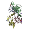
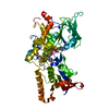
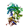
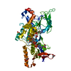
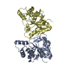
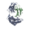
- PDB-5tug: Archaellum periplasmic stator protein complex FlaF and FlaG from ... -
+ Open data

- Basic information
| Entry | Database: PDB / ID: 5tug | ||||||
|---|---|---|---|---|---|---|---|
| Title | Archaellum periplasmic stator protein complex FlaF and FlaG from Sulfolobus acidocaldarius | ||||||
 Components Components | (Flagellar biosynthesis protein ...) x 2 | ||||||
 Keywords Keywords | MOTOR PROTEIN / beta-sandwich fold Archaellum assembly subunit Stator protein | ||||||
| Function / homology |  Function and homology information Function and homology informationarchaeal or bacterial-type flagellum-dependent cell motility / structural molecule activity / membrane / identical protein binding Similarity search - Function | ||||||
| Biological species |   Sulfolobus acidocaldarius (acidophilic) Sulfolobus acidocaldarius (acidophilic) | ||||||
| Method |  X-RAY DIFFRACTION / SYNCHROTRON / MOLECULAR REPLACEMENT / molecular replacement / Resolution: 2.47 Å X-RAY DIFFRACTION / SYNCHROTRON / MOLECULAR REPLACEMENT / molecular replacement / Resolution: 2.47 Å | ||||||
 Authors Authors | Tsai, C.-L. / Tainer, J.A. | ||||||
 Citation Citation | Journal: Nat Microbiol / Year: 2020 Title: The structure of the periplasmic FlaG-FlaF complex and its essential role for archaellar swimming motility. Authors: Tsai, C.L. / Tripp, P. / Sivabalasarma, S. / Zhang, C. / Rodriguez-Franco, M. / Wipfler, R.L. / Chaudhury, P. / Banerjee, A. / Beeby, M. / Whitaker, R.J. / Tainer, J.A. / Albers, S.V. | ||||||
| History |
|
- Structure visualization
| Structure viewer | Molecule: MolmilJmol/JSmol |
|---|
- Downloads & links
-Download
| PDBx/mmCIF format | 5tug.cif.gz | 115.1 KB | Display | PDBx/mmCIF format |
|---|---|---|---|---|
| PDB format | pdb5tug.ent.gz | 87.7 KB | Display | PDB format |
| PDBx/mmJSON format | 5tug.json.gz | Tree view | PDBx/mmJSON format | |
| Others |  Other downloads Other downloads |
-Validation report
| Arichive directory | https://data.pdbj.org/pub/pdb/validation_reports/tu/5tugftp://data.pdbj.org/pub/pdb/validation_reports/tu/5tug | HTTPS FTP |
|---|
-Related structure data
| Related structure data | 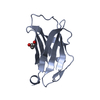 5tuhC  6pbkC 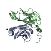 4p94S S: Starting model for refinement C: citing same article ( |
|---|---|
| Similar structure data |


-Links
 PDBj
PDBj


- Assembly
| Deposited unit | 
| ||||||||
|---|---|---|---|---|---|---|---|---|---|
| 1 | 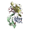
| ||||||||
| Unit cell |
|
-Components
-Flagellar biosynthesis protein ... , 2 types, 4 molecules ACBD
| #1: Protein | Mass: 14941.408 Da / Num. of mol.: 2 / Fragment: UNP residues 32-151 Source method: isolated from a genetically manipulated source Source: (gene. exp.) Sulfolobus acidocaldarius (acidophilic)Gene: ATY89_00610, ATZ20_03655 / Plasmid: pET-Duet1 / Production host:  #2: Protein | Mass: 16248.860 Da / Num. of mol.: 2 / Fragment: UNP residues 35-164 Source method: isolated from a genetically manipulated source Source: (gene. exp.) Sulfolobus acidocaldarius (acidophilic)Gene: ATY89_00615, ATZ20_03660 / Plasmid: pET-Duet1 / Production host: |
|---|
-Non-polymers , 4 types, 196 molecules 


| #3: Chemical | ChemComp-EDO /  Mass: 62.068 Da / Num. of mol.: 5 / Source method: obtained synthetically / Formula: C2H6O2 Mass: 62.068 Da / Num. of mol.: 5 / Source method: obtained synthetically / Formula: C2H6O2#4: Chemical |  Mass: 106.120 Da / Num. of mol.: 3 / Source method: obtained synthetically / Formula: C4H10O3 Mass: 106.120 Da / Num. of mol.: 3 / Source method: obtained synthetically / Formula: C4H10O3#5: Chemical | ChemComp-IMD /  Mass: 69.085 Da / Num. of mol.: 4 / Source method: obtained synthetically / Formula: C3H5N2 Mass: 69.085 Da / Num. of mol.: 4 / Source method: obtained synthetically / Formula: C3H5N2#6: Water | ChemComp-HOH / | Mass: 18.015 Da / Num. of mol.: 184 / Source method: isolated from a natural source / Formula: H2O |
|---|
-Experimental details
-Experiment
| Experiment | Method: X-RAY DIFFRACTION / Number of used crystals: 1 |
|---|
- Sample preparation
| Crystal | Density Matthews: 5.07 Å3/Da / Density % sol: 75.74 % / Description: hexagonal |
|---|---|
| Crystal grow | Temperature: 288 K / Method: vapor diffusion, hanging drop / pH: 7.5 / Details: 18% PEG 6000 0.1M Tris, pH 7.5, 0.2M NaBr |
-Data collection
| Diffraction | Mean temperature: 100 K |
|---|---|
| Diffraction source | Source: SYNCHROTRON / Site: ALS  / Beamline: 12.3.1 / Wavelength: 0.9918 Å / Beamline: 12.3.1 / Wavelength: 0.9918 Å |
| Detector | Type: ADSC QUANTUM 315r / Detector: CCD / Date: Jul 9, 2015 |
| Radiation | Monochromator: ML crystals / Protocol: SINGLE WAVELENGTH / Monochromatic (M) / Laue (L): M / Scattering type: x-ray |
| Radiation wavelength | Wavelength: 0.9918 Å / Relative weight: 1 |
| Reflection twin | Operator: h,-h-k,-l / Fraction: 0.24 |
| Reflection | Resolution: 2.47→49.152 Å / Num. obs: 44562 / % possible obs: 100 % / Redundancy: 11.4 % / CC1/2: 0.999 / Rmerge(I) obs: 0.101 / Net I/σ(I): 19.2 |
| Reflection shell | Resolution: 2.47→2.56 Å / Redundancy: 11.5 % / Rmerge(I) obs: 1.291 / Mean I/σ(I) obs: 2.4 / CC1/2: 0.774 / % possible all: 100 |
-Phasing
| Phasing | Method: molecular replacement |
|---|
- Processing
| Software |
| ||||||||||||||||||||||||||||||||||||||||||||||||||||||||||||||||||||||||||||||||||||||||||||||||||||||||||||||||
|---|---|---|---|---|---|---|---|---|---|---|---|---|---|---|---|---|---|---|---|---|---|---|---|---|---|---|---|---|---|---|---|---|---|---|---|---|---|---|---|---|---|---|---|---|---|---|---|---|---|---|---|---|---|---|---|---|---|---|---|---|---|---|---|---|---|---|---|---|---|---|---|---|---|---|---|---|---|---|---|---|---|---|---|---|---|---|---|---|---|---|---|---|---|---|---|---|---|---|---|---|---|---|---|---|---|---|---|---|---|---|---|---|---|
| Refinement | Method to determine structure: MOLECULAR REPLACEMENT Starting model: 4P94 Resolution: 2.47→49.152 Å / Cross valid method: FREE R-VALUE / σ(F): 1.34 / Phase error: 28.89 / Stereochemistry target values: TWIN_LSQ_F
| ||||||||||||||||||||||||||||||||||||||||||||||||||||||||||||||||||||||||||||||||||||||||||||||||||||||||||||||||
| Solvent computation | Shrinkage radii: 0.9 Å / VDW probe radii: 1.11 Å / Solvent model: FLAT BULK SOLVENT MODEL | ||||||||||||||||||||||||||||||||||||||||||||||||||||||||||||||||||||||||||||||||||||||||||||||||||||||||||||||||
| Refinement step | Cycle: LAST / Resolution: 2.47→49.152 Å
| ||||||||||||||||||||||||||||||||||||||||||||||||||||||||||||||||||||||||||||||||||||||||||||||||||||||||||||||||
| Refine LS restraints |
| ||||||||||||||||||||||||||||||||||||||||||||||||||||||||||||||||||||||||||||||||||||||||||||||||||||||||||||||||
| LS refinement shell |
|
