 Movie
Movie Controller


+ Open data

- Basic information
| Entry | Database: PDB / ID: 5tee | ||||||
|---|---|---|---|---|---|---|---|
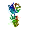
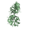
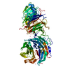
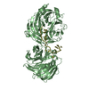
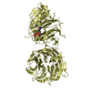
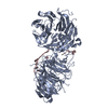
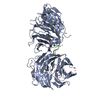
| Title | Crystal structure of Gemin5 WD40 repeats in apo form | ||||||
 Components Components | Gem-associated protein 5 | ||||||
 Keywords Keywords | SPLICING / Structural Genomics / Structural Genomics Consortium / SGC | ||||||
| Function / homology |  Function and homology information Function and homology informationSMN-Gemin2 complex / Gemini of Cajal bodies / SMN complex / U4atac snRNA binding / snRNA binding / RNA 7-methylguanosine cap binding / SMN-Sm protein complex / U4 snRNA binding / spliceosomal snRNP assembly / U1 snRNA binding ...SMN-Gemin2 complex / Gemini of Cajal bodies / SMN complex / U4atac snRNA binding / snRNA binding / RNA 7-methylguanosine cap binding / SMN-Sm protein complex / U4 snRNA binding / spliceosomal snRNP assembly / U1 snRNA binding / mRNA 3'-UTR binding / mRNA splicing, via spliceosome / regulation of translation / ribosome binding / protein-containing complex assembly / snRNP Assembly / SARS-CoV-2 modulates host translation machinery / nuclear body / translation / RNA binding / nucleoplasm / membrane / nucleus / cytoplasm / cytosol Similarity search - Function | ||||||
| Biological species |  Homo sapiens (human) Homo sapiens (human) | ||||||
| Method |  X-RAY DIFFRACTION / SYNCHROTRON / MOLECULAR REPLACEMENT / Resolution: 1.65 Å X-RAY DIFFRACTION / SYNCHROTRON / MOLECULAR REPLACEMENT / Resolution: 1.65 Å | ||||||
 Authors Authors | Chao, X. / Tempel, W. / Bian, C. / Cerovina, T. / He, H. / Walker, J.R. / Seitova, A. / Bountra, C. / Arrowsmith, C.H. / Edwards, A.M. ...Chao, X. / Tempel, W. / Bian, C. / Cerovina, T. / He, H. / Walker, J.R. / Seitova, A. / Bountra, C. / Arrowsmith, C.H. / Edwards, A.M. / Min, J. / Structural Genomics Consortium (SGC) | ||||||
 Citation Citation | Journal: Genes Dev. / Year: 2016 Title: Structural insights into Gemin5-guided selection of pre-snRNAs for snRNP assembly. Authors: Xu, C. / Ishikawa, H. / Izumikawa, K. / Li, L. / He, H. / Nobe, Y. / Yamauchi, Y. / Shahjee, H.M. / Wu, X.H. / Yu, Y.T. / Isobe, T. / Takahashi, N. / Min, J. | ||||||
| History |
|
- Structure visualization
| Structure viewer | Molecule: MolmilJmol/JSmol |
|---|
- Downloads & links
-Download
| PDBx/mmCIF format | 5tee.cif.gz | 166.1 KB | Display | PDBx/mmCIF format |
|---|---|---|---|---|
| PDB format | pdb5tee.ent.gz | 125.6 KB | Display | PDB format |
| PDBx/mmJSON format | 5tee.json.gz | Tree view | PDBx/mmJSON format | |
| Others |  Other downloads Other downloads |
-Validation report
| Arichive directory | https://data.pdbj.org/pub/pdb/validation_reports/te/5teeftp://data.pdbj.org/pub/pdb/validation_reports/te/5tee | HTTPS FTP |
|---|
-Related structure data
| Related structure data | 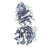 5gxhC  5gxiC  5tefC  5thaC  2hesS 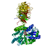 3dm0S  3ow8S C: citing same article ( S: Starting model for refinement |
|---|---|
| Similar structure data |


-Links
 PDBj
PDBj


- Assembly
| Deposited unit | 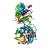
| ||||||||
|---|---|---|---|---|---|---|---|---|---|
| 1 |
| ||||||||
| Unit cell |
| ||||||||
| Details | Authors did not provide a biological assembly |
-Components
| #1: Protein | Mass: 84170.195 Da / Num. of mol.: 1 Source method: isolated from a genetically manipulated source Source: (gene. exp.) Homo sapiens (human) / Gene: GEMIN5 / Plasmid: pFBOH-LIC / Production host:   Spodoptera frugiperda (fall armyworm) / Strain (production host): SF9 / References: UniProt: Q8TEQ6 Spodoptera frugiperda (fall armyworm) / Strain (production host): SF9 / References: UniProt: Q8TEQ6 | ||||||||
|---|---|---|---|---|---|---|---|---|---|
| #2: Chemical |   Mass: 92.094 Da / Num. of mol.: 3 / Source method: obtained synthetically / Formula: C3H8O3 Mass: 92.094 Da / Num. of mol.: 3 / Source method: obtained synthetically / Formula: C3H8O3#3: Chemical | ChemComp-UNX /   Num. of mol.: 35 / Source method: obtained synthetically Num. of mol.: 35 / Source method: obtained synthetically#4: Chemical |   Mass: 22.990 Da / Num. of mol.: 2 / Source method: obtained synthetically / Formula: Na Mass: 22.990 Da / Num. of mol.: 2 / Source method: obtained synthetically / Formula: Na#5: Water | ChemComp-HOH / |  Mass: 18.015 Da / Num. of mol.: 420 / Source method: isolated from a natural source / Formula: H2O Mass: 18.015 Da / Num. of mol.: 420 / Source method: isolated from a natural source / Formula: H2OHas protein modification | Y | |
-Experimental details
-Experiment
| Experiment | Method: X-RAY DIFFRACTION / Number of used crystals: 1 |
|---|
- Sample preparation
| Crystal | Density Matthews: 2.6 Å3/Da / Density % sol: 53.1 % |
|---|---|
| Crystal grow | Temperature: 291 K / Method: vapor diffusion / pH: 5.5 Details: 15% PEG4000, 0.2M ammonium acetate, 0.1M sodium citrate |
-Data collection
| Diffraction | Mean temperature: 100 K | ||||||||||||||||||||||||||||||
|---|---|---|---|---|---|---|---|---|---|---|---|---|---|---|---|---|---|---|---|---|---|---|---|---|---|---|---|---|---|---|---|
| Diffraction source | Source: SYNCHROTRON / Site: APS  / Beamline: 19-ID / Wavelength: 0.97918 Å / Beamline: 19-ID / Wavelength: 0.97918 Å | ||||||||||||||||||||||||||||||
| Detector | Type: ADSC QUANTUM 315r / Detector: CCD / Date: Aug 18, 2011 | ||||||||||||||||||||||||||||||
| Radiation | Protocol: SINGLE WAVELENGTH / Monochromatic (M) / Laue (L): M / Scattering type: x-ray | ||||||||||||||||||||||||||||||
| Radiation wavelength | Wavelength: 0.97918 Å / Relative weight: 1 | ||||||||||||||||||||||||||||||
| Reflection | Resolution: 1.65→48.71 Å / Num. obs: 93484 / % possible obs: 98.9 % / Redundancy: 3.8 % / CC1/2: 0.997 / Rmerge(I) obs: 0.091 / Rpim(I) all: 0.054 / Rrim(I) all: 0.107 / Net I/σ(I): 10.8 / Num. measured all: 355460 | ||||||||||||||||||||||||||||||
| Reflection shell | Diffraction-ID: 1 / Rejects: _
|
- Processing
| Software |
| |||||||||||||||||||||||||||||||||||||||||||||||||||||||||||||||||||||||||||
|---|---|---|---|---|---|---|---|---|---|---|---|---|---|---|---|---|---|---|---|---|---|---|---|---|---|---|---|---|---|---|---|---|---|---|---|---|---|---|---|---|---|---|---|---|---|---|---|---|---|---|---|---|---|---|---|---|---|---|---|---|---|---|---|---|---|---|---|---|---|---|---|---|---|---|---|---|
| Refinement | Method to determine structure: MOLECULAR REPLACEMENT Starting model: FFAS03/SCWRL models based on PDB entries 3ow8, 2hes, 3dm0 Resolution: 1.65→48.47 Å / Cor.coef. Fo:Fc: 0.967 / Cor.coef. Fo:Fc free: 0.952 / SU B: 1.992 / SU ML: 0.064 / Cross valid method: THROUGHOUT / σ(F): 0 / ESU R: 0.084 / ESU R Free: 0.085 Details: Molecular replacement search models were based on PDB entries 3ow8, 2hes, 3dm0 and modified using SCWRL and the FFAS03 server. Programs Phaser and Molrep were used for molecular replacement. ...Details: Molecular replacement search models were based on PDB entries 3ow8, 2hes, 3dm0 and modified using SCWRL and the FFAS03 server. Programs Phaser and Molrep were used for molecular replacement. Arp/warp was used for map imrpovement and automated model building. Geometry restraints for the ligand were prepared with PRODRG based on GTG coordinates from PDB entry 3HXI. Coot was used for interactive model building. PHENIX.molprobity was used for geometry validation.
| |||||||||||||||||||||||||||||||||||||||||||||||||||||||||||||||||||||||||||
| Solvent computation | Ion probe radii: 0.8 Å / Shrinkage radii: 0.8 Å / VDW probe radii: 1.2 Å | |||||||||||||||||||||||||||||||||||||||||||||||||||||||||||||||||||||||||||
| Displacement parameters | Biso max: 82.85 Å2 / Biso mean: 21.435 Å2 / Biso min: 10.11 Å2
| |||||||||||||||||||||||||||||||||||||||||||||||||||||||||||||||||||||||||||
| Refinement step | Cycle: final / Resolution: 1.65→48.47 Å
| |||||||||||||||||||||||||||||||||||||||||||||||||||||||||||||||||||||||||||
| Refine LS restraints |
| |||||||||||||||||||||||||||||||||||||||||||||||||||||||||||||||||||||||||||
| LS refinement shell | Resolution: 1.65→1.693 Å / Total num. of bins used: 20
|
