- PDB-3dm0: Maltose Binding Protein fusion with RACK1 from A. thaliana -
+
Open data
ID or keywords:
Loading...
-
Basic information
Entry
Database: PDB / ID: 3dm0
Title
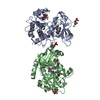
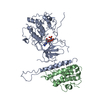
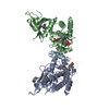
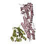
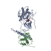
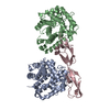
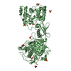
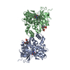
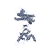
Maltose Binding Protein fusion with RACK1 from A. thaliana
Components
Maltose-binding periplasmic protein fused with RACK1
Keywords
Sugar Binding Protein / Signaling Protein / MBP RACK1A / Receptor for activiated protein C-kinase 1 / beta-propeller WD40 repeat / Sugar transport / Transport / WD repeat
Function / homology
Function and homology information
gibberellin mediated signaling pathway / cellular response to abscisic acid stimulus / response to gibberellin / vegetative to reproductive phase transition of meristem / seed germination / positive regulation of signal transduction / MAP kinase scaffold activity / detection of maltose stimulus / maltose transport complex / carbohydrate transport ...gibberellin mediated signaling pathway / cellular response to abscisic acid stimulus / response to gibberellin / vegetative to reproductive phase transition of meristem / seed germination / positive regulation of signal transduction / MAP kinase scaffold activity / detection of maltose stimulus / maltose transport complex / carbohydrate transport / negative regulation of translational frameshifting / carbohydrate transmembrane transporter activity / maltose binding / maltose transport / maltodextrin transmembrane transport / response to glucose / ATP-binding cassette (ABC) transporter complex, substrate-binding subunit-containing / translation regulator activity / ATP-binding cassette (ABC) transporter complex / rescue of stalled cytosolic ribosome / cytosolic ribosome / protein kinase C binding / cell chemotaxis / chloroplast / regulation of translation / outer membrane-bounded periplasmic space / ribosome binding / ribosome biogenesis / molecular adaptor activity / periplasmic space / structural constituent of ribosome / ribonucleoprotein complex / mRNA binding / DNA damage response / membrane / nucleus / cytoplasm / cytosol Similarity search - Function
Maltose/Cyclodextrin ABC transporter, substrate-binding protein / Solute-binding family 1, conserved site / Bacterial extracellular solute-binding proteins, family 1 signature. / Bacterial extracellular solute-binding protein / Small (40S) ribosomal subunit Asc1/RACK1 / Bacterial extracellular solute-binding protein / YVTN repeat-like/Quinoprotein amine dehydrogenase / 7 Propeller / Methylamine Dehydrogenase; Chain H / Periplasmic binding protein-like II ...Maltose/Cyclodextrin ABC transporter, substrate-binding protein / Solute-binding family 1, conserved site / Bacterial extracellular solute-binding proteins, family 1 signature. / Bacterial extracellular solute-binding protein / Small (40S) ribosomal subunit Asc1/RACK1 / Bacterial extracellular solute-binding protein / YVTN repeat-like/Quinoprotein amine dehydrogenase / 7 Propeller / Methylamine Dehydrogenase; Chain H / Periplasmic binding protein-like II / D-Maltodextrin-Binding Protein; domain 2 / WD domain, G-beta repeat / G-protein beta WD-40 repeat / WD40 repeat, conserved site / Trp-Asp (WD) repeats signature. / Trp-Asp (WD) repeats profile. / Trp-Asp (WD) repeats circular profile. / WD40 repeats / WD40 repeat / WD40-repeat-containing domain superfamily / WD40/YVTN repeat-like-containing domain superfamily / 3-Layer(aba) Sandwich / Mainly Beta / Alpha Beta Similarity search - Domain/homology
alpha-maltose / Small ribosomal subunit protein RACK1z / Maltose/maltodextrin-binding periplasmic protein Similarity search - Component
Biological species
Escherichia coli (E. coli) Arabidopsis thaliana (thale cress)
Method
X-RAY DIFFRACTION / MOLECULAR REPLACEMENT / Resolution: 2.4 Å
In the structure databanks used in Yorodumi, some data are registered as the other names, "COVID-19 virus" and "2019-nCoV". Here are the details of the virus and the list of structure data.
Jan 31, 2019. EMDB accession codes are about to change! (news from PDBe EMDB page)
EMDB accession codes are about to change! (news from PDBe EMDB page)
The allocation of 4 digits for EMDB accession codes will soon come to an end. Whilst these codes will remain in use, new EMDB accession codes will include an additional digit and will expand incrementally as the available range of codes is exhausted. The current 4-digit format prefixed with “EMD-” (i.e. EMD-XXXX) will advance to a 5-digit format (i.e. EMD-XXXXX), and so on. It is currently estimated that the 4-digit codes will be depleted around Spring 2019, at which point the 5-digit format will come into force.
The EM Navigator/Yorodumi systems omit the EMD- prefix.
Related info.:Q: What is EMD? / ID/Accession-code notation in Yorodumi/EM Navigator
Yorodumi is a browser for structure data from EMDB, PDB, SASBDB, etc.
This page is also the successor to EM Navigator detail page, and also detail information page/front-end page for Omokage search.
The word "yorodu" (or yorozu) is an old Japanese word meaning "ten thousand". "mi" (miru) is to see.
Related info.:EMDB / PDB / SASBDB / Comparison of 3 databanks / Yorodumi Search / Aug 31, 2016. New EM Navigator & Yorodumi / Yorodumi Papers / Jmol/JSmol / Function and homology information / Changes in new EM Navigator and Yorodumi
 Movie
Movie Controller


 Open data
 Basic information
 Components
 Keywords
 Function and homology information

 X-RAY DIFFRACTION /
  Authors
 Citation
 Structure visualization
 Downloads & links
 Other downloads
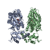

 PDBj
PDBj


 Assembly
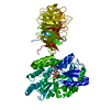


 Mass: 62.068 Da / Num. of mol.: 1 / Source method: obtained synthetically / Formula: C2H6O2
 Mass: 18.015 Da / Num. of mol.: 273 / Source method: isolated from a natural source / Formula: H2O
 Sample preparation
 Processing