 Movie
Movie Controller

[English] 日本語
 Yorodumi
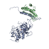
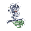
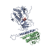
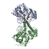
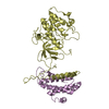
- PDB-4lqp: Crystal structure of the Cbk1(T743E)-Mob2 kinase-coactivator comp... -
+ Open data

- Basic information
| Entry | Database: PDB / ID: 4lqp | ||||||
|---|---|---|---|---|---|---|---|
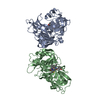
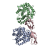
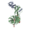
| Title | Crystal structure of the Cbk1(T743E)-Mob2 kinase-coactivator complex, in crystal form A | ||||||
 Components Components |
| ||||||
 Keywords Keywords | TRANSFERASE/TRANSFERASE activator / Kinase / TRANSFERASE-TRANSFERASE activator complex | ||||||
| Function / homology |  Function and homology information Function and homology informationbudding cell apical bud growth / regulation of fungal-type cell wall organization / establishment or maintenance of actin cytoskeleton polarity / prospore membrane / cellular bud / incipient cellular bud site / septum digestion after cytokinesis / cellular bud tip / serine/threonine protein kinase complex / cellular bud neck ...budding cell apical bud growth / regulation of fungal-type cell wall organization / establishment or maintenance of actin cytoskeleton polarity / prospore membrane / cellular bud / incipient cellular bud site / septum digestion after cytokinesis / cellular bud tip / serine/threonine protein kinase complex / cellular bud neck / mating projection tip / regulation of protein secretion / establishment or maintenance of cell polarity / protein kinase activator activity / cytoplasmic stress granule / cell cortex / non-specific serine/threonine protein kinase / intracellular signal transduction / protein serine kinase activity / cell division / protein serine/threonine kinase activity / signal transduction / ATP binding / identical protein binding / nucleus / cytoplasm Similarity search - Function | ||||||
| Biological species |  | ||||||
| Method |  X-RAY DIFFRACTION / SYNCHROTRON / MOLECULAR REPLACEMENT / Resolution: 4.5 Å X-RAY DIFFRACTION / SYNCHROTRON / MOLECULAR REPLACEMENT / Resolution: 4.5 Å | ||||||
 Authors Authors | Gogl, G. / Remenyi, A. | ||||||
 Citation Citation | Journal: Plos Biol. / Year: 2015 Title: The Structure of an NDR/LATS Kinase-Mob Complex Reveals a Novel Kinase-Coactivator System and Substrate Docking Mechanism. Authors: Gogl, G. / Schneider, K.D. / Yeh, B.J. / Alam, N. / Nguyen Ba, A.N. / Moses, A.M. / Hetenyi, C. / Remenyi, A. / Weiss, E.L. | ||||||
| History |
|
- Structure visualization
| Structure viewer | Molecule: MolmilJmol/JSmol |
|---|
- Downloads & links
-Download
| PDBx/mmCIF format | 4lqp.cif.gz | 128.2 KB | Display | PDBx/mmCIF format |
|---|---|---|---|---|
| PDB format | pdb4lqp.ent.gz | 93.4 KB | Display | PDB format |
| PDBx/mmJSON format | 4lqp.json.gz | Tree view | PDBx/mmJSON format | |
| Others |  Other downloads Other downloads |
-Validation report
| Arichive directory | https://data.pdbj.org/pub/pdb/validation_reports/lq/4lqpftp://data.pdbj.org/pub/pdb/validation_reports/lq/4lqp | HTTPS FTP |
|---|
-Related structure data


-Links
 PDBj
PDBj


- Assembly
| Deposited unit | 
| ||||||||
|---|---|---|---|---|---|---|---|---|---|
| 1 |
| ||||||||
| Unit cell |
|
-Components
| #1: Protein | Mass: 58784.582 Da / Num. of mol.: 1 / Fragment: UNP residues 251-756 / Mutation: D475A, T743E Source method: isolated from a genetically manipulated source Source: (gene. exp.) Strain: ATCC 204508 / S288c / Gene: CBK1, N1727, YNL161W / Production host:  References: UniProt: P53894, non-specific serine/threonine protein kinase |
|---|---|
| #2: Protein | Mass: 28415.197 Da / Num. of mol.: 1 / Fragment: UNP residues 46-287 Source method: isolated from a genetically manipulated source Source: (gene. exp.) Strain: ATCC 204508 / S288c / Gene: MOB2, YFL034C-B, YFL035C, YFL035C-A / Production host: |
| #3: Chemical | ChemComp-ANP /   Mass: 506.196 Da / Num. of mol.: 1 / Source method: obtained synthetically / Formula: C10H17N6O12P3 / Comment: AMP-PNP, energy-carrying molecule analogue*YM Mass: 506.196 Da / Num. of mol.: 1 / Source method: obtained synthetically / Formula: C10H17N6O12P3 / Comment: AMP-PNP, energy-carrying molecule analogue*YM |
-Experimental details
-Experiment
| Experiment | Method: X-RAY DIFFRACTION / Number of used crystals: 1 |
|---|
- Sample preparation
| Crystal | Density Matthews: 5.56 Å3/Da / Density % sol: 77.88 % |
|---|---|
| Crystal grow | Temperature: 296 K / Method: microbatch under oil / pH: 5.5 Details: 25% PEG 20,000 buffered with 0.1M Na-citrate, pH 5.5, Microbatch under oil, temperature 296K |
-Data collection
| Diffraction | Mean temperature: 100 K |
|---|---|
| Diffraction source | Source: SYNCHROTRON / Site: SLS  / Beamline: X06SA / Wavelength: 1 Å / Beamline: X06SA / Wavelength: 1 Å |
| Detector | Type: PSI PILATUS 6M / Detector: PIXEL / Date: May 28, 2011 |
| Radiation | Monochromator: SI(111) / Protocol: SINGLE WAVELENGTH / Monochromatic (M) / Laue (L): M / Scattering type: x-ray |
| Radiation wavelength | Wavelength: 1 Å / Relative weight: 1 |
| Reflection | Resolution: 4.5→49.591 Å / Num. all: 12243 / Num. obs: 12238 / % possible obs: 98.9 % / Observed criterion σ(F): 2.83 / Observed criterion σ(I): 2.83 |
| Reflection shell | Resolution: 4.5→4.66 Å / % possible all: 100 |
- Processing
| Software |
| |||||||||||||||||||||||||||||||||||
|---|---|---|---|---|---|---|---|---|---|---|---|---|---|---|---|---|---|---|---|---|---|---|---|---|---|---|---|---|---|---|---|---|---|---|---|---|
| Refinement | Method to determine structure: MOLECULAR REPLACEMENT / Resolution: 4.5→49.59 Å / SU ML: 0.6 / σ(F): 1.35 / Phase error: 39.29 / Stereochemistry target values: ML
| |||||||||||||||||||||||||||||||||||
| Solvent computation | Shrinkage radii: 0.9 Å / VDW probe radii: 1.11 Å / Solvent model: FLAT BULK SOLVENT MODEL | |||||||||||||||||||||||||||||||||||
| Refinement step | Cycle: LAST / Resolution: 4.5→49.59 Å
| |||||||||||||||||||||||||||||||||||
| Refine LS restraints |
| |||||||||||||||||||||||||||||||||||
| LS refinement shell |
|
