 Movie
Movie Controller


+ Open data

- Basic information
| Entry | Database: PDB / ID: 5ta0 | |||||||||
|---|---|---|---|---|---|---|---|---|---|---|
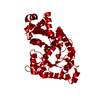
| Title | Crystal structure of BuGH86E322Q in complex with neoagarooctaose | |||||||||
 Components Components | Glycoside Hydrolase | |||||||||
 Keywords Keywords | HYDROLASE / (alpha/beta)6 barrel / glycoside hydrolase | |||||||||
| Function / homology |  Function and homology information Function and homology informationJelly Rolls - #1200 / Porphyranase catalytic subdomain 1 / Beta-porphyranase A, C-terminal / beta porphyranase A C-terminal / Porphyranase beta-sandwich domain 1 / Glycoside hydrolase superfamily / Jelly Rolls / Sandwich / Mainly Beta Similarity search - Domain/homology | |||||||||
| Biological species |  Bacteroides uniformis (bacteria) Bacteroides uniformis (bacteria) | |||||||||
| Method |  X-RAY DIFFRACTION / SYNCHROTRON / MOLECULAR REPLACEMENT / molecular replacement / Resolution: 1.4 Å X-RAY DIFFRACTION / SYNCHROTRON / MOLECULAR REPLACEMENT / molecular replacement / Resolution: 1.4 Å | |||||||||
 Authors Authors | Pluvinage, B. / Boraston, A.B. / Abbott, W.D. | |||||||||
 Citation Citation | Journal: Nat Commun / Year: 2018 Title: Molecular basis of an agarose metabolic pathway acquired by a human intestinal symbiont. Authors: Pluvinage, B. / Grondin, J.M. / Amundsen, C. / Klassen, L. / Moote, P.E. / Xiao, Y. / Thomas, D. / Pudlo, N.A. / Anele, A. / Martens, E.C. / Inglis, G.D. / Uwiera, R.E.R. / Boraston, A.B. / Abbott, D.W. | |||||||||
| History |
|
- Structure visualization
| Structure viewer | Molecule: MolmilJmol/JSmol |
|---|
- Downloads & links
-Download
| PDBx/mmCIF format | 5ta0.cif.gz | 312.1 KB | Display | PDBx/mmCIF format |
|---|---|---|---|---|
| PDB format | pdb5ta0.ent.gz | 248.2 KB | Display | PDB format |
| PDBx/mmJSON format | 5ta0.json.gz | Tree view | PDBx/mmJSON format | |
| Others |  Other downloads Other downloads |
-Validation report
| Arichive directory | https://data.pdbj.org/pub/pdb/validation_reports/ta/5ta0ftp://data.pdbj.org/pub/pdb/validation_reports/ta/5ta0 | HTTPS FTP |
|---|
-Related structure data
| Related structure data |  5t98C  5t99C  5t9aC  5t9gC 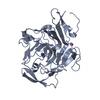 5t9xC 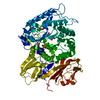 5ta1C 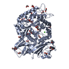 5ta5C  5ta7C  5ta9C C: citing same article ( |
|---|---|
| Similar structure data |


-Links
 PDBj
PDBj- Assembly
| Deposited unit | 
| ||||||||
|---|---|---|---|---|---|---|---|---|---|
| 1 | 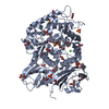
| ||||||||
| 2 | 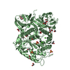
| ||||||||
| 3 | 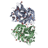
| ||||||||
| Unit cell |
|
-Components
-Protein / Sugars , 2 types, 3 molecules AB
| #1: Protein | Mass: 74747.688 Da / Num. of mol.: 2 / Mutation: E322Q Source method: isolated from a genetically manipulated source Source: (gene. exp.) Bacteroides uniformis (bacteria) / Strain: NP1 / Plasmid: pET28a / Production host: #2: Polysaccharide | beta-D-galactopyranose-(1-4)-3,6-anhydro-alpha-L-galactopyranose-(1-3)-beta-D-galactopyranose-(1-4)- ...beta-D-galactopyranose-(1-4)-3,6-anhydro-alpha-L-galactopyranose-(1-3)-beta-D-galactopyranose-(1-4)-3,6-anhydro-alpha-L-galactopyranose-(1-3)-beta-D-galactopyranose | Source method: isolated from a genetically manipulated source |
|---|
-Non-polymers , 5 types, 1363 molecules 


| #3: Chemical | ChemComp-SO4 /  Mass: 96.063 Da / Num. of mol.: 8 / Source method: obtained synthetically / Formula: SO4 Mass: 96.063 Da / Num. of mol.: 8 / Source method: obtained synthetically / Formula: SO4#4: Chemical | ChemComp-EDO /  Mass: 62.068 Da / Num. of mol.: 30 / Source method: obtained synthetically / Formula: C2H6O2 Mass: 62.068 Da / Num. of mol.: 30 / Source method: obtained synthetically / Formula: C2H6O2#5: Chemical | ChemComp-GOL /  Mass: 92.094 Da / Num. of mol.: 9 / Source method: obtained synthetically / Formula: C3H8O3 Mass: 92.094 Da / Num. of mol.: 9 / Source method: obtained synthetically / Formula: C3H8O3#6: Chemical | ChemComp-CA /  Mass: 40.078 Da / Num. of mol.: 4 / Source method: obtained synthetically / Formula: Ca Mass: 40.078 Da / Num. of mol.: 4 / Source method: obtained synthetically / Formula: Ca#7: Water | ChemComp-HOH / | Mass: 18.015 Da / Num. of mol.: 1312 / Source method: isolated from a natural source / Formula: H2O |
|---|
-Details
| Has protein modification | Y |
|---|
-Experimental details
-Experiment
| Experiment | Method: X-RAY DIFFRACTION / Number of used crystals: 1 |
|---|
- Sample preparation
| Crystal | Density Matthews: 2.35 Å3/Da / Density % sol: 47.72 % |
|---|---|
| Crystal grow | Temperature: 291 K / Method: vapor diffusion, hanging drop / pH: 6.2 Details: 0.16M CaOAc, 0.1M Na cacodylate, 17.5% PEG 8000, 5% glycerol |
-Data collection
| Diffraction | Mean temperature: 100 K |
|---|---|
| Diffraction source | Source: SYNCHROTRON / Site: CLSI  / Beamline: 08ID-1 / Wavelength: 0.984 Å / Beamline: 08ID-1 / Wavelength: 0.984 Å |
| Detector | Type: RAYONIX MX-300 / Detector: CCD / Date: Mar 18, 2015 |
| Radiation | Protocol: SINGLE WAVELENGTH / Monochromatic (M) / Laue (L): M / Scattering type: x-ray |
| Radiation wavelength | Wavelength: 0.984 Å / Relative weight: 1 |
| Reflection | Resolution: 1.4→85.9 Å / Num. obs: 256936 / % possible obs: 95.7 % / Redundancy: 6.6 % / CC1/2: 0.998 / Rmerge(I) obs: 0.07 / Net I/σ(I): 15.7 |
| Reflection shell | Resolution: 1.4→1.48 Å / Redundancy: 6.6 % / Rmerge(I) obs: 0.324 / Mean I/σ(I) obs: 5.4 / CC1/2: 0.952 / % possible all: 93.4 |
-Phasing
| Phasing | Method: molecular replacement |
|---|
- Processing
| Software |
| |||||||||||||||||||||||||||||||||||||||||||||||||||||||||||||||||||||||||||
|---|---|---|---|---|---|---|---|---|---|---|---|---|---|---|---|---|---|---|---|---|---|---|---|---|---|---|---|---|---|---|---|---|---|---|---|---|---|---|---|---|---|---|---|---|---|---|---|---|---|---|---|---|---|---|---|---|---|---|---|---|---|---|---|---|---|---|---|---|---|---|---|---|---|---|---|---|
| Refinement | Method to determine structure: MOLECULAR REPLACEMENT / Resolution: 1.4→82.88 Å / Cor.coef. Fo:Fc: 0.974 / Cor.coef. Fo:Fc free: 0.967 / SU B: 0.76 / SU ML: 0.031 / Cross valid method: THROUGHOUT / σ(F): 0 / ESU R: 0.052 / ESU R Free: 0.053 Details: HYDROGENS HAVE BEEN ADDED IN THE RIDING POSITIONS U VALUES : REFINED INDIVIDUALLY
| |||||||||||||||||||||||||||||||||||||||||||||||||||||||||||||||||||||||||||
| Solvent computation | Ion probe radii: 0.8 Å / Shrinkage radii: 0.8 Å / VDW probe radii: 1.2 Å | |||||||||||||||||||||||||||||||||||||||||||||||||||||||||||||||||||||||||||
| Displacement parameters | Biso max: 70.9 Å2 / Biso mean: 13.685 Å2 / Biso min: 4.76 Å2
| |||||||||||||||||||||||||||||||||||||||||||||||||||||||||||||||||||||||||||
| Refinement step | Cycle: final / Resolution: 1.4→82.88 Å
| |||||||||||||||||||||||||||||||||||||||||||||||||||||||||||||||||||||||||||
| Refine LS restraints |
| |||||||||||||||||||||||||||||||||||||||||||||||||||||||||||||||||||||||||||
| LS refinement shell | Resolution: 1.4→1.436 Å / Total num. of bins used: 20
|
