 Movie
Movie Controller

[English] 日本語
 Yorodumi
- PDB-7cm8: High resolution crystal structure of M92A mutant of O-acetyl-L-se... -
+ Open data

- Basic information
| Entry | Database: PDB / ID: 7cm8 | ||||||
|---|---|---|---|---|---|---|---|
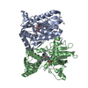
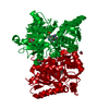
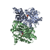
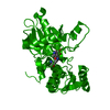
| Title | High resolution crystal structure of M92A mutant of O-acetyl-L-serine sulfhydrylase from Haemophilus influenzae | ||||||
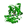
 Components Components | Cysteine synthase | ||||||
 Keywords Keywords | TRANSFERASE / Cysteine synthase / Substrate selection / mutant / Competetive allostery | ||||||
| Function / homology |  Function and homology information Function and homology informationL-cysteine desulfhydrase activity / cysteine synthase / cysteine synthase activity / : / cytoplasm Similarity search - Function | ||||||
| Biological species |  Haemophilus influenzae (bacteria) Haemophilus influenzae (bacteria) | ||||||
| Method |  X-RAY DIFFRACTION / MOLECULAR REPLACEMENT / Resolution: 1.9 Å X-RAY DIFFRACTION / MOLECULAR REPLACEMENT / Resolution: 1.9 Å | ||||||
 Authors Authors | Kaushik, A. / Rahisuddin, R. / Saini, N. / Kumaran, S. | ||||||
 Citation Citation | Journal: J.Biol.Chem. / Year: 2020 Title: Molecular mechanism of selective substrate engagement and inhibitor disengagement of cysteine synthase. Authors: Kaushik, A. / Rahisuddin, R. / Saini, N. / Singh, R.P. / Kaur, R. / Koul, S. / Kumaran, S. | ||||||
| History |
|
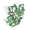
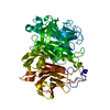
- Structure visualization
| Structure viewer | Molecule: MolmilJmol/JSmol |
|---|
- Downloads & links
-Download
| PDBx/mmCIF format | 7cm8.cif.gz | 77.1 KB | Display | PDBx/mmCIF format |
|---|---|---|---|---|
| PDB format | pdb7cm8.ent.gz | 53 KB | Display | PDB format |
| PDBx/mmJSON format | 7cm8.json.gz | Tree view | PDBx/mmJSON format | |
| Others |  Other downloads Other downloads |
-Validation report
| Arichive directory | https://data.pdbj.org/pub/pdb/validation_reports/cm/7cm8ftp://data.pdbj.org/pub/pdb/validation_reports/cm/7cm8 | HTTPS FTP |
|---|
-Related structure data
| Related structure data | 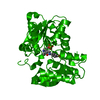 5xcnC  5xcpSC 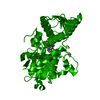 5xcwC  7c35C S: Starting model for refinement C: citing same article ( |
|---|---|
| Similar structure data |


-Links
 PDBj
PDBj
- Assembly
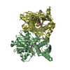
| Deposited unit | 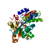
| ||||||||
|---|---|---|---|---|---|---|---|---|---|
| 1 | 
| ||||||||
| Unit cell |
|
-Components
| #1: Protein | Mass: 37144.273 Da / Num. of mol.: 1 / Mutation: M92A Source method: isolated from a genetically manipulated source Source: (gene. exp.) Haemophilus influenzae (strain ATCC 51907 / DSM 11121 / KW20 / Rd) (bacteria)Gene: cysK, HI_1103 / Plasmid: peT28a+ PBR322 Production host: References: UniProt: P45040, cysteine synthase | ||||||
|---|---|---|---|---|---|---|---|
| #2: Chemical | ChemComp-NA /   Mass: 22.990 Da / Num. of mol.: 5 / Source method: obtained synthetically / Formula: Na / Feature type: SUBJECT OF INVESTIGATION Mass: 22.990 Da / Num. of mol.: 5 / Source method: obtained synthetically / Formula: Na / Feature type: SUBJECT OF INVESTIGATION#3: Chemical | ChemComp-GOL / |   Mass: 92.094 Da / Num. of mol.: 1 / Source method: obtained synthetically / Formula: C3H8O3 / Feature type: SUBJECT OF INVESTIGATION Mass: 92.094 Da / Num. of mol.: 1 / Source method: obtained synthetically / Formula: C3H8O3 / Feature type: SUBJECT OF INVESTIGATION#4: Water | ChemComp-HOH / |  Mass: 18.015 Da / Num. of mol.: 77 / Source method: obtained synthetically / Formula: H2O Mass: 18.015 Da / Num. of mol.: 77 / Source method: obtained synthetically / Formula: H2OHas ligand of interest | Y | |
-Experimental details
-Experiment
| Experiment | Method: X-RAY DIFFRACTION / Number of used crystals: 1 |
|---|
- Sample preparation
| Crystal | Density Matthews: 1.88 Å3/Da / Density % sol: 34.7 % |
|---|---|
| Crystal grow | Temperature: 291 K / Method: vapor diffusion, sitting drop / pH: 7.4 / Details: 100mM HEPES buffer pH 7.4, 1.4M Sodium Citrate |
-Data collection
| Diffraction | Mean temperature: 100 K / Serial crystal experiment: N | ||||||||||||||||||
|---|---|---|---|---|---|---|---|---|---|---|---|---|---|---|---|---|---|---|---|
| Diffraction source | Source: ROTATING ANODE / Type: RIGAKU MICROMAX-007 HF / Wavelength: 1.5418 Å | ||||||||||||||||||
| Detector | Type: MAR scanner 345 mm plate / Detector: IMAGE PLATE / Date: Jul 7, 2020 | ||||||||||||||||||
| Radiation | Monochromator: M / Protocol: SINGLE WAVELENGTH / Monochromatic (M) / Laue (L): M / Scattering type: x-ray | ||||||||||||||||||
| Radiation wavelength | Wavelength: 1.5418 Å / Relative weight: 1 | ||||||||||||||||||
| Reflection twin |
| ||||||||||||||||||
| Reflection | Resolution: 1.9→39.75 Å / Num. obs: 21690 / % possible obs: 98.9 % / Redundancy: 10.2 % / Biso Wilson estimate: 21.66 Å2 / Rmerge(I) obs: 0.145 / Net I/σ(I): 9.6 | ||||||||||||||||||
| Reflection shell | Resolution: 1.9→1.97 Å / Redundancy: 9.9 % / Rmerge(I) obs: 0.82 / Mean I/σ(I) obs: 2.7 / Num. unique obs: 1380 / % possible all: 98.6 |
- Processing
| Software |
| ||||||||||||||||||||
|---|---|---|---|---|---|---|---|---|---|---|---|---|---|---|---|---|---|---|---|---|---|
| Refinement | Method to determine structure: MOLECULAR REPLACEMENT Starting model: 5XCP Resolution: 1.9→35.56 Å / Cor.coef. Fo:Fc: 0.95 / Cor.coef. Fo:Fc free: 0.943 / SU B: 4.053 / SU ML: 0.119 / Cross valid method: THROUGHOUT / ESU R: 0.028 / ESU R Free: 0.032 / Stereochemistry target values: MAXIMUM LIKELIHOOD / Details: HYDROGENS HAVE BEEN ADDED IN THE RIDING POSITIONS
| ||||||||||||||||||||
| Solvent computation | Ion probe radii: 0.8 Å / Shrinkage radii: 0.8 Å / VDW probe radii: 1.2 Å / Solvent model: MASK | ||||||||||||||||||||
| Displacement parameters | Biso mean: 32.031 Å2
| ||||||||||||||||||||
| Refinement step | Cycle: 1 / Resolution: 1.9→35.56 Å
| ||||||||||||||||||||
| LS refinement shell | Resolution: 1.9→1.95 Å / Total num. of bins used: 20
|
