 Movie
Movie Controller

[English] 日本語
 Yorodumi
- PDB-7djq: Crystal Structure of O-acetyl L-serine sulfhydrylase from Haemoph... -
+ Open data

- Basic information
| Entry | Database: PDB / ID: 7djq | ||||||
|---|---|---|---|---|---|---|---|
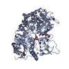
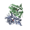
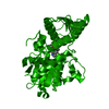
| Title | Crystal Structure of O-acetyl L-serine sulfhydrylase from Haemophilus influenzae in complex with C-Terminal peptide of ribosomal S4 Domain protein from Lactobacillus salivarius. | ||||||
 Components Components |
| ||||||
 Keywords Keywords | TRANSFERASE / Complex / Enzyme / inhibitor | ||||||
| Function / homology |  Function and homology information Function and homology informationL-cysteine desulfhydrase activity / cysteine synthase / cysteine synthase activity / : / RNA binding / cytoplasm Similarity search - Function | ||||||
| Biological species |  Haemophilus influenzae (bacteria)Lactobacillus salivarius UCC118 (bacteria) Haemophilus influenzae (bacteria)Lactobacillus salivarius UCC118 (bacteria) | ||||||
| Method |  X-RAY DIFFRACTION / MOLECULAR REPLACEMENT / Resolution: 2.3 Å X-RAY DIFFRACTION / MOLECULAR REPLACEMENT / Resolution: 2.3 Å | ||||||
 Authors Authors | Saini, N. / Rahisuddin, R. / Kumaran, S. | ||||||
 Citation Citation | Journal: J.Mol.Biol. / Year: 2021 Title: Moonlighting Biochemistry of Cysteine Synthase: A Species-specific Global Regulator. Authors: Singh, R.P. / Saini, N. / Sharma, G. / Rahisuddin, R. / Patel, M. / Kaushik, A. / Kumaran, S. | ||||||
| History |
|
- Structure visualization
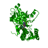
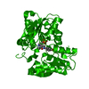
| Structure viewer | Molecule: MolmilJmol/JSmol |
|---|
- Downloads & links
-Download
| PDBx/mmCIF format | 7djq.cif.gz | 220.6 KB | Display | PDBx/mmCIF format |
|---|---|---|---|---|
| PDB format | pdb7djq.ent.gz | 173.9 KB | Display | PDB format |
| PDBx/mmJSON format | 7djq.json.gz | Tree view | PDBx/mmJSON format | |
| Others |  Other downloads Other downloads |
-Validation report
| Arichive directory | https://data.pdbj.org/pub/pdb/validation_reports/dj/7djqftp://data.pdbj.org/pub/pdb/validation_reports/dj/7djq | HTTPS FTP |
|---|
-Related structure data
| Related structure data | 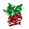 4ho1S S: Starting model for refinement |
|---|---|
| Similar structure data |


-Links
 PDBj
PDBj
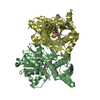
- Assembly
| Deposited unit | 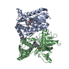
| ||||||||||||
|---|---|---|---|---|---|---|---|---|---|---|---|---|---|
| 1 |
| ||||||||||||
| Unit cell |
|
-Components
| #1: Protein/peptide | Mass: 1188.221 Da / Num. of mol.: 1 / Source method: obtained synthetically / Source: (synth.) Lactobacillus salivarius UCC118 (bacteria) / References: UniProt: A0A1V9TQZ2 | ||||||
|---|---|---|---|---|---|---|---|
| #2: Protein | Mass: 37244.453 Da / Num. of mol.: 2 / Mutation: D67E/A68P Source method: isolated from a genetically manipulated source Source: (gene. exp.) Haemophilus influenzae (strain ATCC 51907 / DSM 11121 / KW20 / Rd) (bacteria)Gene: cysK, HI_1103 Production host: References: UniProt: P45040, cysteine synthase #3: Chemical |   Mass: 22.990 Da / Num. of mol.: 3 / Source method: obtained synthetically / Formula: Na / Feature type: SUBJECT OF INVESTIGATION Mass: 22.990 Da / Num. of mol.: 3 / Source method: obtained synthetically / Formula: Na / Feature type: SUBJECT OF INVESTIGATION#4: Water | ChemComp-HOH / |  Mass: 18.015 Da / Num. of mol.: 125 / Source method: isolated from a natural source / Formula: H2O Mass: 18.015 Da / Num. of mol.: 125 / Source method: isolated from a natural source / Formula: H2OHas ligand of interest | Y | |
-Experimental details
-Experiment
| Experiment | Method: X-RAY DIFFRACTION / Number of used crystals: 1 |
|---|
- Sample preparation
| Crystal | Density Matthews: 2.33 Å3/Da / Density % sol: 47.1 % |
|---|---|
| Crystal grow | Temperature: 291 K / Method: vapor diffusion, sitting drop / pH: 7.4 / Details: 100mM HEPES buffer pH 7.4, 1.4M Sodium Citrate |
-Data collection
| Diffraction | Mean temperature: 100 K / Serial crystal experiment: N |
|---|---|
| Diffraction source | Source: ROTATING ANODE / Type: RIGAKU MICROMAX-007 HF / Wavelength: 1.5418 Å |
| Detector | Type: MAR scanner 345 mm plate / Detector: IMAGE PLATE / Date: Mar 19, 2020 |
| Radiation | Monochromator: M / Protocol: SINGLE WAVELENGTH / Monochromatic (M) / Laue (L): M / Scattering type: x-ray |
| Radiation wavelength | Wavelength: 1.5418 Å / Relative weight: 1 |
| Reflection | Resolution: 2.3→40.86 Å / Num. obs: 28307 / % possible obs: 97.66 % / Redundancy: 15.2 % / Biso Wilson estimate: 19.86 Å2 / CC1/2: 0.855 / CC star: 0.96 / Rmerge(I) obs: 0.063 / Net I/σ(I): 17.1 |
| Reflection shell | Resolution: 2.3→2.382 Å / Redundancy: 9 % / Rmerge(I) obs: 0.15 / Num. unique obs: 2705 / CC1/2: 0.826 / CC star: 0.951 / % possible all: 94.98 |
- Processing
| Software |
| |||||||||||||||||||||||||||||||||||||||||||||||||||||||||||||||||||||||||||||
|---|---|---|---|---|---|---|---|---|---|---|---|---|---|---|---|---|---|---|---|---|---|---|---|---|---|---|---|---|---|---|---|---|---|---|---|---|---|---|---|---|---|---|---|---|---|---|---|---|---|---|---|---|---|---|---|---|---|---|---|---|---|---|---|---|---|---|---|---|---|---|---|---|---|---|---|---|---|---|
| Refinement | Method to determine structure: MOLECULAR REPLACEMENT Starting model: 4HO1 Resolution: 2.3→40.86 Å / SU ML: 0.2184 / Cross valid method: FREE R-VALUE / σ(F): 1.35 / Phase error: 23.1909 / Stereochemistry target values: CDL v1.2
| |||||||||||||||||||||||||||||||||||||||||||||||||||||||||||||||||||||||||||||
| Solvent computation | Shrinkage radii: 0.9 Å / VDW probe radii: 1.11 Å / Solvent model: FLAT BULK SOLVENT MODEL | |||||||||||||||||||||||||||||||||||||||||||||||||||||||||||||||||||||||||||||
| Displacement parameters | Biso mean: 22.76 Å2 | |||||||||||||||||||||||||||||||||||||||||||||||||||||||||||||||||||||||||||||
| Refinement step | Cycle: LAST / Resolution: 2.3→40.86 Å
| |||||||||||||||||||||||||||||||||||||||||||||||||||||||||||||||||||||||||||||
| Refine LS restraints |
| |||||||||||||||||||||||||||||||||||||||||||||||||||||||||||||||||||||||||||||
| LS refinement shell |
|
