 Movie
Movie Controller

[English] 日本語
 Yorodumi
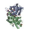
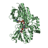
- PDB-5v9h: Structure of the H477R variant of rat cytosolic PEPCK in complex ... -
+ Open data

- Basic information
| Entry | Database: PDB / ID: 5v9h | ||||||
|---|---|---|---|---|---|---|---|
| Title | Structure of the H477R variant of rat cytosolic PEPCK in complex with phosphoglycolate and GDP. | ||||||
 Components Components | Phosphoenolpyruvate carboxykinase, cytosolic [GTP] | ||||||
 Keywords Keywords | LYASE / phosphoenolpyruvate carboxykinase | ||||||
| Function / homology |  Function and homology information Function and homology informationresponse to methionine / Gluconeogenesis / phosphoenolpyruvate carboxykinase activity / protein serine kinase activity (using GTP as donor) / SREBP-SCAP complex retention in endoplasmic reticulum / Transferases; Transferring phosphorus-containing groups; Protein-serine/threonine kinases / cellular response to potassium ion starvation / phosphoenolpyruvate carboxykinase (GTP) / phosphoenolpyruvate carboxykinase (GTP) activity / : ...response to methionine / Gluconeogenesis / phosphoenolpyruvate carboxykinase activity / protein serine kinase activity (using GTP as donor) / SREBP-SCAP complex retention in endoplasmic reticulum / Transferases; Transferring phosphorus-containing groups; Protein-serine/threonine kinases / cellular response to potassium ion starvation / phosphoenolpyruvate carboxykinase (GTP) / phosphoenolpyruvate carboxykinase (GTP) activity / : / propionate catabolic process / cellular response to raffinose / tricarboxylic acid metabolic process / response to interleukin-6 / regulation of lipid biosynthetic process / cellular response to fructose stimulus / cellular hypotonic salinity response / cellular hypotonic response / carboxylic acid binding / cellular response to phorbol 13-acetate 12-myristate / oxaloacetate metabolic process / hepatocyte differentiation / positive regulation of memory T cell differentiation / cellular hyperosmotic response / glyceraldehyde-3-phosphate biosynthetic process / nucleoside diphosphate kinase activity / response to acidic pH / NLS-bearing protein import into nucleus / cellular hyperosmotic salinity response / response to starvation / response to lipid / cellular response to dexamethasone stimulus / cellular response to interleukin-1 / positive regulation of lipid biosynthetic process / cellular response to retinoic acid / cellular response to glucagon stimulus / response to bacterium / cellular response to cAMP / response to activity / gluconeogenesis / peptidyl-serine phosphorylation / lipid metabolic process / cellular response to glucose stimulus / protein maturation / response to insulin / response to nutrient levels / positive regulation of cholesterol biosynthetic process / glucose metabolic process / cellular response to tumor necrosis factor / cellular response to insulin stimulus / GDP binding / glucose homeostasis / manganese ion binding / response to lipopolysaccharide / cellular response to hypoxia / GTP binding / magnesium ion binding / endoplasmic reticulum / positive regulation of transcription by RNA polymerase II / mitochondrion / cytosol / cytoplasm Similarity search - Function | ||||||
| Biological species |  | ||||||
| Method |  X-RAY DIFFRACTION / MOLECULAR REPLACEMENT / molecular replacement / Resolution: 2.15 Å X-RAY DIFFRACTION / MOLECULAR REPLACEMENT / molecular replacement / Resolution: 2.15 Å | ||||||
 Authors Authors | Holyoak, T. / Cui, D.S. | ||||||
| Funding support |  Canada, 1items Canada, 1items
| ||||||
 Citation Citation | Journal: Biochemistry / Year: 2017 Title: Asymmetric Anchoring Is Required for Efficient Omega-Loop Opening and Closing in Cytosolic Phosphoenolpyruvate Carboxykinase. Authors: Cui, D.S. / Broom, A. / Mcleod, M.J. / Meiering, E.M. / Holyoak, T. | ||||||
| History |
|
- Structure visualization
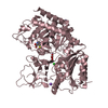
| Structure viewer | Molecule: MolmilJmol/JSmol |
|---|
- Downloads & links
-Download
| PDBx/mmCIF format | 5v9h.cif.gz | 260 KB | Display | PDBx/mmCIF format |
|---|---|---|---|---|
| PDB format | pdb5v9h.ent.gz | 204.3 KB | Display | PDB format |
| PDBx/mmJSON format | 5v9h.json.gz | Tree view | PDBx/mmJSON format | |
| Others |  Other downloads Other downloads |
-Validation report
| Arichive directory | https://data.pdbj.org/pub/pdb/validation_reports/v9/5v9hftp://data.pdbj.org/pub/pdb/validation_reports/v9/5v9h | HTTPS FTP |
|---|
-Related structure data
| Related structure data | 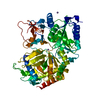 5v95C 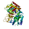 5v97C 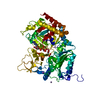 5v9fC 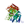 5v9gC 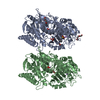 3dtbS C: citing same article ( S: Starting model for refinement |
|---|---|
| Similar structure data |


-Links
 PDBj
PDBj
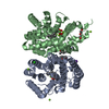
- Assembly
| Deposited unit | 
| ||||||||
|---|---|---|---|---|---|---|---|---|---|
| 1 | 
| ||||||||
| 2 | 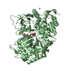
| ||||||||
| Unit cell |
|
-Components
-Protein , 1 types, 2 molecules AB
| #1: Protein | Mass: 69518.711 Da / Num. of mol.: 2 / Mutation: H477R Source method: isolated from a genetically manipulated source Source: (gene. exp.)  References: UniProt: P07379, phosphoenolpyruvate carboxykinase (GTP) |
|---|
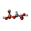
-Non-polymers , 6 types, 303 molecules 


| #2: Chemical | ChemComp-1PE /  Mass: 238.278 Da / Num. of mol.: 1 / Source method: obtained synthetically / Formula: C10H22O6 / Comment: precipitant*YM Mass: 238.278 Da / Num. of mol.: 1 / Source method: obtained synthetically / Formula: C10H22O6 / Comment: precipitant*YM | ||||||||
|---|---|---|---|---|---|---|---|---|---|
| #3: Chemical | ChemComp-MN /  Mass: 54.938 Da / Num. of mol.: 6 / Source method: obtained synthetically / Formula: Mn Mass: 54.938 Da / Num. of mol.: 6 / Source method: obtained synthetically / Formula: Mn#4: Chemical |  Mass: 22.990 Da / Num. of mol.: 2 / Source method: obtained synthetically / Formula: Na Mass: 22.990 Da / Num. of mol.: 2 / Source method: obtained synthetically / Formula: Na#5: Chemical |  Type: RNA linking / Mass: 443.201 Da / Num. of mol.: 2 / Source method: obtained synthetically / Formula: C10H15N5O11P2 / Comment: GDP, energy-carrying molecule*YM Type: RNA linking / Mass: 443.201 Da / Num. of mol.: 2 / Source method: obtained synthetically / Formula: C10H15N5O11P2 / Comment: GDP, energy-carrying molecule*YM#6: Chemical |  Mass: 156.031 Da / Num. of mol.: 2 Mass: 156.031 Da / Num. of mol.: 2Source method: isolated from a genetically manipulated source Formula: C2H5O6P #7: Water | ChemComp-HOH / | Mass: 18.015 Da / Num. of mol.: 290 / Source method: isolated from a natural source / Formula: H2O |
-Experimental details
-Experiment
| Experiment | Method: X-RAY DIFFRACTION / Number of used crystals: 1 |
|---|
- Sample preparation
| Crystal | Density Matthews: 2.21 Å3/Da / Density % sol: 44.32 % / Description: rods |
|---|---|
| Crystal grow | Temperature: 298 K / Method: vapor diffusion, hanging drop / pH: 7.4 / Details: 24 - 34% PEG 3350 and 100mM HEPES, at pH 7.4 |
-Data collection
| Diffraction | Mean temperature: 100 K | |||||||||||||||||||||||||||||||||||||||||||||||||||||||||||||||||||||||||||||||||||||||||||||||||||
|---|---|---|---|---|---|---|---|---|---|---|---|---|---|---|---|---|---|---|---|---|---|---|---|---|---|---|---|---|---|---|---|---|---|---|---|---|---|---|---|---|---|---|---|---|---|---|---|---|---|---|---|---|---|---|---|---|---|---|---|---|---|---|---|---|---|---|---|---|---|---|---|---|---|---|---|---|---|---|---|---|---|---|---|---|---|---|---|---|---|---|---|---|---|---|---|---|---|---|---|---|
| Diffraction source | Source: ROTATING ANODE / Type: RIGAKU / Wavelength: 1.54 Å | |||||||||||||||||||||||||||||||||||||||||||||||||||||||||||||||||||||||||||||||||||||||||||||||||||
| Detector | Type: RIGAKU RAXIS IV++ / Detector: IMAGE PLATE / Date: Aug 17, 2013 | |||||||||||||||||||||||||||||||||||||||||||||||||||||||||||||||||||||||||||||||||||||||||||||||||||
| Radiation | Protocol: SINGLE WAVELENGTH / Monochromatic (M) / Laue (L): M / Scattering type: x-ray | |||||||||||||||||||||||||||||||||||||||||||||||||||||||||||||||||||||||||||||||||||||||||||||||||||
| Radiation wavelength | Wavelength: 1.54 Å / Relative weight: 1 | |||||||||||||||||||||||||||||||||||||||||||||||||||||||||||||||||||||||||||||||||||||||||||||||||||
| Reflection | Resolution: 2.15→100 Å / Num. obs: 65917 / % possible obs: 99.9 % / Redundancy: 7 % / Rmerge(I) obs: 0.132 / Rpim(I) all: 0.054 / Rrim(I) all: 0.143 / Χ2: 1.074 / Net I/σ(I): 10.3 / Num. measured all: 458307 | |||||||||||||||||||||||||||||||||||||||||||||||||||||||||||||||||||||||||||||||||||||||||||||||||||
| Reflection shell | Diffraction-ID: 1
|
-Phasing
| Phasing | Method: molecular replacement |
|---|
- Processing
| Software |
| ||||||||||||||||||||||||||||||||||||||||||||||||||||||||||||
|---|---|---|---|---|---|---|---|---|---|---|---|---|---|---|---|---|---|---|---|---|---|---|---|---|---|---|---|---|---|---|---|---|---|---|---|---|---|---|---|---|---|---|---|---|---|---|---|---|---|---|---|---|---|---|---|---|---|---|---|---|---|
| Refinement | Method to determine structure: MOLECULAR REPLACEMENT Starting model: 3DTB Resolution: 2.15→85.04 Å / Cor.coef. Fo:Fc: 0.952 / Cor.coef. Fo:Fc free: 0.931 / SU B: 10.772 / SU ML: 0.256 / Cross valid method: THROUGHOUT / σ(F): 0 / ESU R: 0.351 / ESU R Free: 0.249 Details: HYDROGENS HAVE BEEN ADDED IN THE RIDING POSITIONS U VALUES : REFINED INDIVIDUALLY
| ||||||||||||||||||||||||||||||||||||||||||||||||||||||||||||
| Solvent computation | Ion probe radii: 0.8 Å / Shrinkage radii: 0.8 Å / VDW probe radii: 1.2 Å | ||||||||||||||||||||||||||||||||||||||||||||||||||||||||||||
| Displacement parameters | Biso max: 114.7 Å2 / Biso mean: 46.667 Å2 / Biso min: 24.52 Å2
| ||||||||||||||||||||||||||||||||||||||||||||||||||||||||||||
| Refinement step | Cycle: final / Resolution: 2.15→85.04 Å
| ||||||||||||||||||||||||||||||||||||||||||||||||||||||||||||
| Refine LS restraints |
| ||||||||||||||||||||||||||||||||||||||||||||||||||||||||||||
| LS refinement shell | Resolution: 2.137→2.192 Å / Rfactor Rfree error: 0 / Total num. of bins used: 20
|
