 Movie
Movie Controller


+ Open data

- Basic information
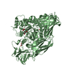
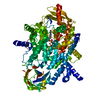
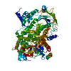
| Entry | Database: PDB / ID: 5t9a | ||||||
|---|---|---|---|---|---|---|---|
| Title | Crystal structure of BuGH2Cwt | ||||||
 Components Components | Glycoside Hydrolase | ||||||
 Keywords Keywords | HYDROLASE / (alpha/beta)6 barrel / glycoside hydrolase | ||||||
| Function / homology |  Function and homology information Function and homology informationhydrolase activity, hydrolyzing O-glycosyl compounds / carbohydrate metabolic process Similarity search - Function | ||||||
| Biological species |  Bacteroides uniformis (bacteria) Bacteroides uniformis (bacteria) | ||||||
| Method |  X-RAY DIFFRACTION / SYNCHROTRON / MOLECULAR REPLACEMENT / molecular replacement / Resolution: 2.5 Å X-RAY DIFFRACTION / SYNCHROTRON / MOLECULAR REPLACEMENT / molecular replacement / Resolution: 2.5 Å | ||||||
 Authors Authors | Pluvinage, B. / Boraston, A.B. / Abbott, W.D. | ||||||
 Citation Citation | Journal: Nat Commun / Year: 2018 Title: Molecular basis of an agarose metabolic pathway acquired by a human intestinal symbiont. Authors: Pluvinage, B. / Grondin, J.M. / Amundsen, C. / Klassen, L. / Moote, P.E. / Xiao, Y. / Thomas, D. / Pudlo, N.A. / Anele, A. / Martens, E.C. / Inglis, G.D. / Uwiera, R.E.R. / Boraston, A.B. / Abbott, D.W. | ||||||
| History |
|
- Structure visualization
| Structure viewer | Molecule: MolmilJmol/JSmol |
|---|
- Downloads & links
-Download
| PDBx/mmCIF format | 5t9a.cif.gz | 656.8 KB | Display | PDBx/mmCIF format |
|---|---|---|---|---|
| PDB format | pdb5t9a.ent.gz | 530.6 KB | Display | PDB format |
| PDBx/mmJSON format | 5t9a.json.gz | Tree view | PDBx/mmJSON format | |
| Others |  Other downloads Other downloads |
-Validation report
| Arichive directory | https://data.pdbj.org/pub/pdb/validation_reports/t9/5t9aftp://data.pdbj.org/pub/pdb/validation_reports/t9/5t9a | HTTPS FTP |
|---|
-Related structure data
| Related structure data | 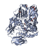 5t98C 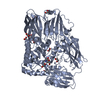 5t99C 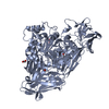 5t9gC 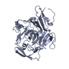 5t9xC 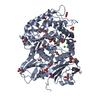 5ta0C 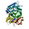 5ta1C 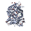 5ta5C  5ta7C  5ta9C 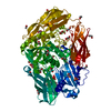 4cu6S C: citing same article ( S: Starting model for refinement |
|---|---|
| Similar structure data |


-Links
 PDBj
PDBj

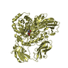
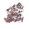
- Assembly
| Deposited unit | 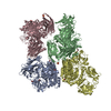
| ||||||||
|---|---|---|---|---|---|---|---|---|---|
| 1 | 
| ||||||||
| 2 | 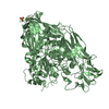
| ||||||||
| 3 | 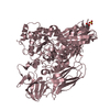
| ||||||||
| 4 | 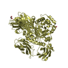
| ||||||||
| Unit cell |
|
-Components
| #1: Protein | Mass: 96870.695 Da / Num. of mol.: 4 Source method: isolated from a genetically manipulated source Source: (gene. exp.) Bacteroides uniformis (bacteria) / Strain: NP1 / Plasmid: pET28a / Production host: #2: Chemical | ChemComp-EDO /   Mass: 62.068 Da / Num. of mol.: 7 / Source method: obtained synthetically / Formula: C2H6O2 Mass: 62.068 Da / Num. of mol.: 7 / Source method: obtained synthetically / Formula: C2H6O2#3: Chemical |   Mass: 96.063 Da / Num. of mol.: 2 / Source method: obtained synthetically / Formula: SO4 Mass: 96.063 Da / Num. of mol.: 2 / Source method: obtained synthetically / Formula: SO4#4: Water | ChemComp-HOH / |  Mass: 18.015 Da / Num. of mol.: 755 / Source method: isolated from a natural source / Formula: H2O Mass: 18.015 Da / Num. of mol.: 755 / Source method: isolated from a natural source / Formula: H2O |
|---|
-Experimental details
-Experiment
| Experiment | Method: X-RAY DIFFRACTION / Number of used crystals: 1 |
|---|
- Sample preparation
| Crystal | Density Matthews: 2.72 Å3/Da / Density % sol: 54.73 % |
|---|---|
| Crystal grow | Temperature: 291 K / Method: vapor diffusion, hanging drop / pH: 8.5 Details: 0.2 M magnesium sulfate, 0.1 M Tris-HCl, 20 % PEG 3350 |
-Data collection
| Diffraction | Mean temperature: 100 K |
|---|---|
| Diffraction source | Source: SYNCHROTRON / Site: CLSI  / Beamline: 08ID-1 / Wavelength: 1 Å / Beamline: 08ID-1 / Wavelength: 1 Å |
| Detector | Type: RAYONIX MX-300 / Detector: CCD / Date: Mar 15, 2015 |
| Radiation | Protocol: SINGLE WAVELENGTH / Monochromatic (M) / Laue (L): M / Scattering type: x-ray |
| Radiation wavelength | Wavelength: 1 Å / Relative weight: 1 |
| Reflection | Resolution: 2.5→117.64 Å / Num. obs: 137926 / % possible obs: 97.8 % / Redundancy: 2.2 % / CC1/2: 0.935 / Rmerge(I) obs: 0.121 / Net I/σ(I): 5.7 |
| Reflection shell | Resolution: 2.5→2.64 Å / Redundancy: 2.2 % / Rmerge(I) obs: 0.548 / CC1/2: 0.298 / % possible all: 97.1 |
-Phasing
| Phasing | Method: molecular replacement |
|---|
- Processing
| Software |
| |||||||||||||||||||||||||||||||||||||||||||||||||||||||||||||||||||||||||||
|---|---|---|---|---|---|---|---|---|---|---|---|---|---|---|---|---|---|---|---|---|---|---|---|---|---|---|---|---|---|---|---|---|---|---|---|---|---|---|---|---|---|---|---|---|---|---|---|---|---|---|---|---|---|---|---|---|---|---|---|---|---|---|---|---|---|---|---|---|---|---|---|---|---|---|---|---|
| Refinement | Method to determine structure: MOLECULAR REPLACEMENT Starting model: 4CU6 Resolution: 2.5→117.64 Å / Cor.coef. Fo:Fc: 0.914 / Cor.coef. Fo:Fc free: 0.864 / SU B: 12.359 / SU ML: 0.264 / Cross valid method: THROUGHOUT / σ(F): 0 / ESU R: 0.887 / ESU R Free: 0.33 Details: HYDROGENS HAVE BEEN ADDED IN THE RIDING POSITIONS U VALUES : REFINED INDIVIDUALLY
| |||||||||||||||||||||||||||||||||||||||||||||||||||||||||||||||||||||||||||
| Solvent computation | Ion probe radii: 0.8 Å / Shrinkage radii: 0.8 Å / VDW probe radii: 1.2 Å | |||||||||||||||||||||||||||||||||||||||||||||||||||||||||||||||||||||||||||
| Displacement parameters | Biso max: 83.36 Å2 / Biso mean: 30.07 Å2 / Biso min: 4.38 Å2
| |||||||||||||||||||||||||||||||||||||||||||||||||||||||||||||||||||||||||||
| Refinement step | Cycle: final / Resolution: 2.5→117.64 Å
| |||||||||||||||||||||||||||||||||||||||||||||||||||||||||||||||||||||||||||
| Refine LS restraints |
| |||||||||||||||||||||||||||||||||||||||||||||||||||||||||||||||||||||||||||
| LS refinement shell | Resolution: 2.5→2.565 Å / Total num. of bins used: 20
|
