 Movie
Movie Controller

[English] 日本語
 Yorodumi
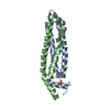
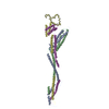
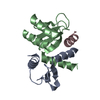
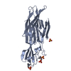
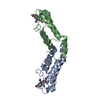
- PDB-5t59: Structure of the MIND Complex Shows a Regulatory Focus of Yeast K... -
+ Open data

- Basic information
| Entry | Database: PDB / ID: 5t59 | ||||||
|---|---|---|---|---|---|---|---|
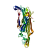
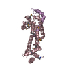
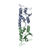
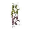
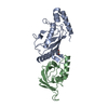
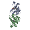
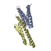
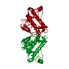
| Title | Structure of the MIND Complex Shows a Regulatory Focus of Yeast Kinetochore Assembly | ||||||
 Components Components |
| ||||||
 Keywords Keywords | CELL CYCLE / kinetochore / complex / chromosome / segregation / MIND / Mis12 | ||||||
| Function / homology |  Function and homology information Function and homology informationMIS12/MIND type complex / spindle attachment to meiosis I kinetochore / centromeric DNA binding / attachment of mitotic spindle microtubules to kinetochore / kinetochore assembly / mitotic sister chromatid segregation / chromosome segregation / kinetochore / cell division / nucleus Similarity search - Function | ||||||
| Biological species |  Kluyveromyces lactis (yeast) Kluyveromyces lactis (yeast) | ||||||
| Method |  X-RAY DIFFRACTION / SYNCHROTRON / SAD / Resolution: 2.405 Å X-RAY DIFFRACTION / SYNCHROTRON / SAD / Resolution: 2.405 Å | ||||||
 Authors Authors | Dimitrova, Y. / Jenni, S. / Valverde, R. / Khin, Y. / Harrison, S.C. | ||||||
 Citation Citation | Journal: Cell / Year: 2016 Title: Structure of the MIND Complex Defines a Regulatory Focus for Yeast Kinetochore Assembly. Authors: Dimitrova, Y.N. / Jenni, S. / Valverde, R. / Khin, Y. / Harrison, S.C. | ||||||
| History |
|
- Structure visualization
| Structure viewer | Molecule: MolmilJmol/JSmol |
|---|
- Downloads & links
-Download
| PDBx/mmCIF format | 5t59.cif.gz | 190.7 KB | Display | PDBx/mmCIF format |
|---|---|---|---|---|
| PDB format | pdb5t59.ent.gz | 154.7 KB | Display | PDB format |
| PDBx/mmJSON format | 5t59.json.gz | Tree view | PDBx/mmJSON format | |
| Others |  Other downloads Other downloads |
-Validation report
| Arichive directory | https://data.pdbj.org/pub/pdb/validation_reports/t5/5t59ftp://data.pdbj.org/pub/pdb/validation_reports/t5/5t59 | HTTPS FTP |
|---|
-Related structure data
-Links
 PDBj
PDBj- Assembly
| Deposited unit | 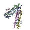
| ||||||||
|---|---|---|---|---|---|---|---|---|---|
| 1 | 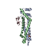
| ||||||||
| 2 | 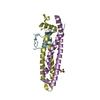
| ||||||||
| Unit cell |
| ||||||||
| Details | Trimer confirmed by gel filtration |
-Components
| #1: Protein | Mass: 13870.916 Da / Num. of mol.: 2 Source method: isolated from a genetically manipulated source Details: Residues Asn0 and Ala1 are left over from the TEV cleavage site. Mtw1 starts at residue Thr2. Source: (gene. exp.) Kluyveromyces lactis (strain ATCC 8585 / CBS 2359 / DSM 70799 / NBRC 1267 / NRRL Y-1140 / WM37) (yeast)Strain: ATCC 8585 / CBS 2359 / DSM 70799 / NBRC 1267 / NRRL Y-1140 / WM37 Gene: KLLA0_F02343g / Production host:  #2: Protein | Mass: 12100.684 Da / Num. of mol.: 2 Source method: isolated from a genetically manipulated source Source: (gene. exp.) Kluyveromyces lactis (strain ATCC 8585 / CBS 2359 / DSM 70799 / NBRC 1267 / NRRL Y-1140 / WM37) (yeast)Strain: ATCC 8585 / CBS 2359 / DSM 70799 / NBRC 1267 / NRRL Y-1140 / WM37 Gene: KLLA0_E05809g / Production host: #3: Protein/peptide | Mass: 4826.354 Da / Num. of mol.: 2 Source method: isolated from a genetically manipulated source Source: (gene. exp.) Kluyveromyces lactis (strain ATCC 8585 / CBS 2359 / DSM 70799 / NBRC 1267 / NRRL Y-1140 / WM37) (yeast)Strain: ATCC 8585 / CBS 2359 / DSM 70799 / NBRC 1267 / NRRL Y-1140 / WM37 Gene: KLLA0_B13629g / Production host: #4: Chemical |   Mass: 207.290 Da / Num. of mol.: 2 / Source method: obtained synthetically / Formula: C8H17NO3S / Comment: pH buffer*YM Mass: 207.290 Da / Num. of mol.: 2 / Source method: obtained synthetically / Formula: C8H17NO3S / Comment: pH buffer*YM#5: Water | ChemComp-HOH / |  Mass: 18.015 Da / Num. of mol.: 33 / Source method: isolated from a natural source / Formula: H2O Mass: 18.015 Da / Num. of mol.: 33 / Source method: isolated from a natural source / Formula: H2O |
|---|
-Experimental details
-Experiment
| Experiment | Method: X-RAY DIFFRACTION / Number of used crystals: 1 |
|---|
- Sample preparation
| Crystal | Density Matthews: 2.68 Å3/Da / Density % sol: 54.14 % |
|---|---|
| Crystal grow | Temperature: 291 K / Method: vapor diffusion, hanging drop / Details: 0.1 M CHES, pH 9.0, 18-30 % PEG 3000 |
-Data collection
| Diffraction | Mean temperature: 100 K |
|---|---|
| Diffraction source | Source: SYNCHROTRON / Site: APS  / Beamline: 24-ID-C / Wavelength: 0.9792 Å / Beamline: 24-ID-C / Wavelength: 0.9792 Å |
| Detector | Type: DECTRIS PILATUS 6M-F / Detector: PIXEL / Date: Oct 27, 2015 |
| Radiation | Protocol: SINGLE WAVELENGTH / Monochromatic (M) / Laue (L): M / Scattering type: x-ray |
| Radiation wavelength | Wavelength: 0.9792 Å / Relative weight: 1 |
| Reflection | Resolution: 2.405→66.17 Å / Num. obs: 16798 / % possible obs: 56.5 % / Redundancy: 3.6 % / CC1/2: 1 / Rmerge(I) obs: 0.073 / Net I/σ(I): 11.9 |
- Processing
| Software |
| |||||||||||||||||||||||||||||||||||||||||||||||||
|---|---|---|---|---|---|---|---|---|---|---|---|---|---|---|---|---|---|---|---|---|---|---|---|---|---|---|---|---|---|---|---|---|---|---|---|---|---|---|---|---|---|---|---|---|---|---|---|---|---|---|
| Refinement | Method to determine structure: SAD / Resolution: 2.405→66.168 Å / SU ML: 0.33 / Cross valid method: FREE R-VALUE / σ(F): 1.37 / Phase error: 36.39
| |||||||||||||||||||||||||||||||||||||||||||||||||
| Solvent computation | Shrinkage radii: 0.6 Å / VDW probe radii: 0.9 Å | |||||||||||||||||||||||||||||||||||||||||||||||||
| Refinement step | Cycle: LAST / Resolution: 2.405→66.168 Å
| |||||||||||||||||||||||||||||||||||||||||||||||||
| Refine LS restraints |
| |||||||||||||||||||||||||||||||||||||||||||||||||
| LS refinement shell |
|
