 ムービー
ムービー コントローラー


+ データを開く

- 基本情報
基本情報
| 登録情報 | データベース: PDB / ID: 5t2s | ||||||
|---|---|---|---|---|---|---|---|
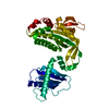
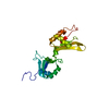
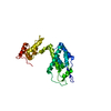
| タイトル | Structure of the FHA1 domain of Rad53 bound simultaneously to the BRCT domain of Dbf4 and a phosphopeptide. | ||||||
 要素 要素 |
| ||||||
 キーワード キーワード | CELL CYCLE / FHA / BRCT / phosphopeptide / protein chimera | ||||||
| 機能・相同性 |  機能・相同性情報 機能・相同性情報positive regulation of spindle attachment to meiosis I kinetochore / positive regulation of meiotic DNA double-strand break formation involved in reciprocal meiotic recombination / positive regulation of kinetochore assembly / positive regulation of meiotic DNA double-strand break formation / positive regulation of DNA replication initiation / negative regulation of exit from mitosis / Dbf4-dependent protein kinase complex / deoxyribonucleoside triphosphate biosynthetic process / positive regulation of protein localization to kinetochore / positive regulation of meiosis I ...positive regulation of spindle attachment to meiosis I kinetochore / positive regulation of meiotic DNA double-strand break formation involved in reciprocal meiotic recombination / positive regulation of kinetochore assembly / positive regulation of meiotic DNA double-strand break formation / positive regulation of DNA replication initiation / negative regulation of exit from mitosis / Dbf4-dependent protein kinase complex / deoxyribonucleoside triphosphate biosynthetic process / positive regulation of protein localization to kinetochore / positive regulation of meiosis I / positive regulation of nuclear cell cycle DNA replication / meiotic recombination checkpoint signaling / regulation of cell cycle phase transition / mitotic DNA replication preinitiation complex assembly / premeiotic DNA replication / Activation of the pre-replicative complex / Activation of ATR in response to replication stress / protein-containing complex localization / dual-specificity kinase / mitotic DNA replication checkpoint signaling / double-strand break repair via break-induced replication / telomere maintenance in response to DNA damage / negative regulation of DNA damage checkpoint / DNA replication origin binding / chromosome, centromeric region / DNA replication initiation / regulation of DNA repair / protein serine/threonine/tyrosine kinase activity / DNA damage checkpoint signaling / protein serine/threonine kinase activator activity / chromosome segregation / intracellular protein localization / protein tyrosine kinase activity / non-specific serine/threonine protein kinase / protein kinase activity / cell division / protein serine kinase activity / DNA repair / protein serine/threonine kinase activity / centrosome / chromatin / signal transduction / zinc ion binding / ATP binding / metal ion binding / identical protein binding / nucleus / cytoplasm / cytosol 類似検索 - 分子機能 | ||||||
| 生物種 |  | ||||||
| 手法 |  X線回折 / シンクロトロン / 分子置換 / 解像度: 2.4 Å X線回折 / シンクロトロン / 分子置換 / 解像度: 2.4 Å | ||||||
 データ登録者 データ登録者 | Guarne, A. / Almawi, A. / Matthews, L. | ||||||
| 資金援助 |  カナダ, 1件 カナダ, 1件
| ||||||
 引用 引用 | ジャーナル: Sci Rep / 年: 2016 タイトル: 'AND' logic gates at work: Crystal structure of Rad53 bound to Dbf4 and Cdc7. 著者: Almawi, A.W. / Matthews, L.A. / Myrox, P. / Boulton, S. / Lai, C. / Moraes, T. / Melacini, G. / Ghirlando, R. / Duncker, B.P. / Guarne, A. | ||||||
| 履歴 |
|
- 構造の表示
構造の表示
| 構造ビューア | 分子: MolmilJmol/JSmol |
|---|
- ダウンロードとリンク
-ダウンロード
| PDBx/mmCIF形式 | 5t2s.cif.gz | 120.7 KB | 表示 | PDBx/mmCIF形式 |
|---|---|---|---|---|
| PDB形式 | pdb5t2s.ent.gz | 91.9 KB | 表示 | PDB形式 |
| PDBx/mmJSON形式 | 5t2s.json.gz | ツリー表示 | PDBx/mmJSON形式 | |
| その他 |  その他のダウンロード その他のダウンロード |
-検証レポート
| 文書・要旨 | 5t2s_validation.pdf.gz | 457.8 KB | 表示 | wwPDB検証レポート |
|---|---|---|---|---|
| 文書・詳細版 | 5t2s_full_validation.pdf.gz | 459.5 KB | 表示 | |
| XML形式データ | 5t2s_validation.xml.gz | 21.7 KB | 表示 | |
| CIF形式データ | 5t2s_validation.cif.gz | 30.5 KB | 表示 | |
| アーカイブディレクトリ | https://data.pdbj.org/pub/pdb/validation_reports/t2/5t2sftp://data.pdbj.org/pub/pdb/validation_reports/t2/5t2s | HTTPS FTP |
-関連構造データ


-リンク
 PDBj
PDBj


- 集合体
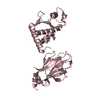
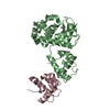
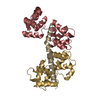
集合体
| 登録構造単位 | 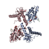
| ||||||||
|---|---|---|---|---|---|---|---|---|---|
| 1 | 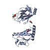
| ||||||||
| 2 | 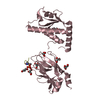
| ||||||||
| 3 |
| ||||||||
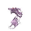
| 単位格子 |
|
-要素
| #1: タンパク質 | 分子量: 30571.002 Da / 分子数: 2 断片: UNP P32325 residues 105-220 linked to UNP P22216 residues 22-162 via LINKER residues VDGS 由来タイプ: 組換発現 由来: (組換発現) 株: ATCC 204508 / S288c 遺伝子: DBF4, DNA52, YDR052C, D4205, YD9609.07C, RAD53, MEC2, SAD1, SPK1, YPL153C, P2588 プラスミド: modified pET15b 詳細 (発現宿主): Contains aTEV-removable His6-SUMO tag 発現宿主:  参照: UniProt: P32325, UniProt: P22216, dual-specificity kinase #2: タンパク質・ペプチド | 分子量: 1347.147 Da / 分子数: 2 / 由来タイプ: 合成 / 詳細: short phosphorylated peptide derived from Cdc7 / 由来: (合成) #3: 化合物 | ChemComp-GOL /   分子量: 92.094 Da / 分子数: 11 / 由来タイプ: 合成 / 式: C3H8O3 分子量: 92.094 Da / 分子数: 11 / 由来タイプ: 合成 / 式: C3H8O3#4: 水 | ChemComp-HOH / |  分子量: 18.015 Da / 分子数: 177 / 由来タイプ: 天然 / 式: H2O 分子量: 18.015 Da / 分子数: 177 / 由来タイプ: 天然 / 式: H2OHas protein modification | Y | |
|---|
-実験情報
-実験
| 実験 | 手法: X線回折 / 使用した結晶の数: 1 |
|---|
- 試料調製
試料調製
| 結晶 | マシュー密度: 2.91 Å3/Da / 溶媒含有率: 57.73 % |
|---|---|
| 結晶化 | 温度: 277 K / 手法: 蒸気拡散法, シッティングドロップ法 / pH: 8.5 / 詳細: 100 mM TRIS pH 8.5 12.5 % PEG 3350 (v/v) / PH範囲: 8.5 |
-データ収集
| 回折 | 平均測定温度: 100 K |
|---|---|
| 放射光源 | 由来: シンクロトロン / サイト: CLSI / ビームライン: 08B1-1 / 波長: 0.979 Å |
| 検出器 | タイプ: RAYONIX MX-300 / 検出器: CCD / 日付: 2015年6月4日 / 詳細: mirrors |
| 放射 | プロトコル: SINGLE WAVELENGTH / 単色(M)・ラウエ(L): M / 散乱光タイプ: x-ray |
| 放射波長 | 波長: 0.979 Å / 相対比: 1 |
| 反射 | 解像度: 2.25→39.674 Å / Num. obs: 36580 / % possible obs: 98.3 % / 冗長度: 2.8 % / CC1/2: 0.99 / Net I/σ(I): 8.15 |
| 反射 シェル | 解像度: 2.2→2.6 Å / 冗長度: 2.8 % / CC1/2: 0.318 / % possible all: 97.4 |
- 解析
解析
| ソフトウェア |
| ||||||||||||||||||||||||||||||||||||||||||||||||||||||||||||||||||||||||||||||||||||
|---|---|---|---|---|---|---|---|---|---|---|---|---|---|---|---|---|---|---|---|---|---|---|---|---|---|---|---|---|---|---|---|---|---|---|---|---|---|---|---|---|---|---|---|---|---|---|---|---|---|---|---|---|---|---|---|---|---|---|---|---|---|---|---|---|---|---|---|---|---|---|---|---|---|---|---|---|---|---|---|---|---|---|---|---|---|
| 精密化 | 構造決定の手法: 分子置換 開始モデル: 1G6G; 3QBZ 解像度: 2.4→39.674 Å / SU ML: 0.32 / 交差検証法: THROUGHOUT / σ(F): 1.36 / 位相誤差: 25.4
| ||||||||||||||||||||||||||||||||||||||||||||||||||||||||||||||||||||||||||||||||||||
| 溶媒の処理 | 減衰半径: 0.9 Å / VDWプローブ半径: 1.11 Å | ||||||||||||||||||||||||||||||||||||||||||||||||||||||||||||||||||||||||||||||||||||
| 原子変位パラメータ | Biso mean: 51.5 Å2 | ||||||||||||||||||||||||||||||||||||||||||||||||||||||||||||||||||||||||||||||||||||
| 精密化ステップ | サイクル: LAST / 解像度: 2.4→39.674 Å
| ||||||||||||||||||||||||||||||||||||||||||||||||||||||||||||||||||||||||||||||||||||
| 拘束条件 |
| ||||||||||||||||||||||||||||||||||||||||||||||||||||||||||||||||||||||||||||||||||||
| LS精密化 シェル |
|
